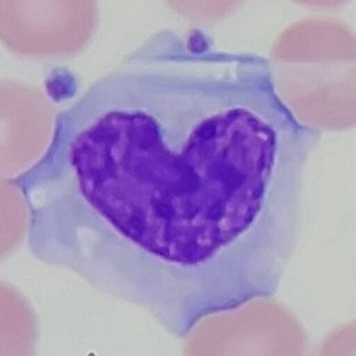
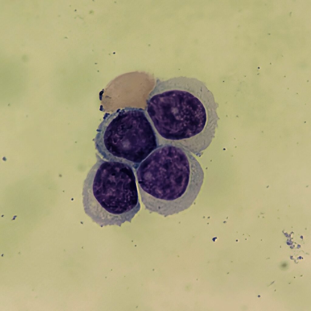
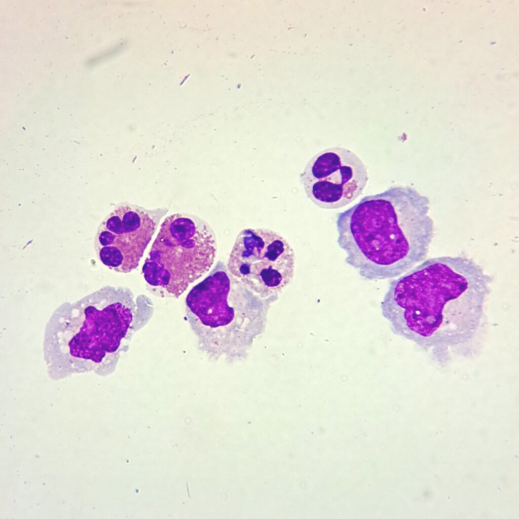
Neutrophils
Cytocentrifugation may make granules in neutrophils appear larger which may be confused for eosinophils. However, they will not have the refractile quality seen in eosinophils or the vibrant reddish-orange coloring.
Large numbers of neutrophils may indicate bacterial infection. In CSF, it is indicative of bacterial meningitis.
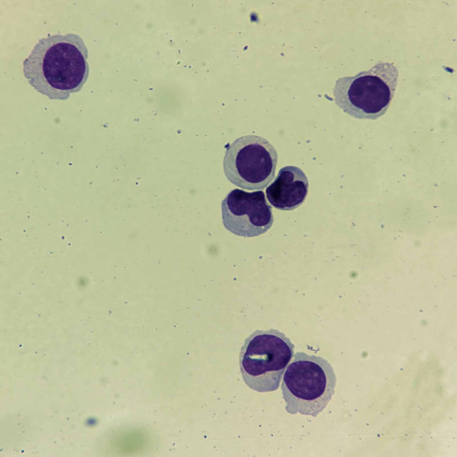
Lymphocytes
Cytocentrifugation may make lymphocytes appear larger which can be mistaken for blasts if not careful.
Increased numbers of lymphs in CSF are indicative of viral meningitis.
More Images
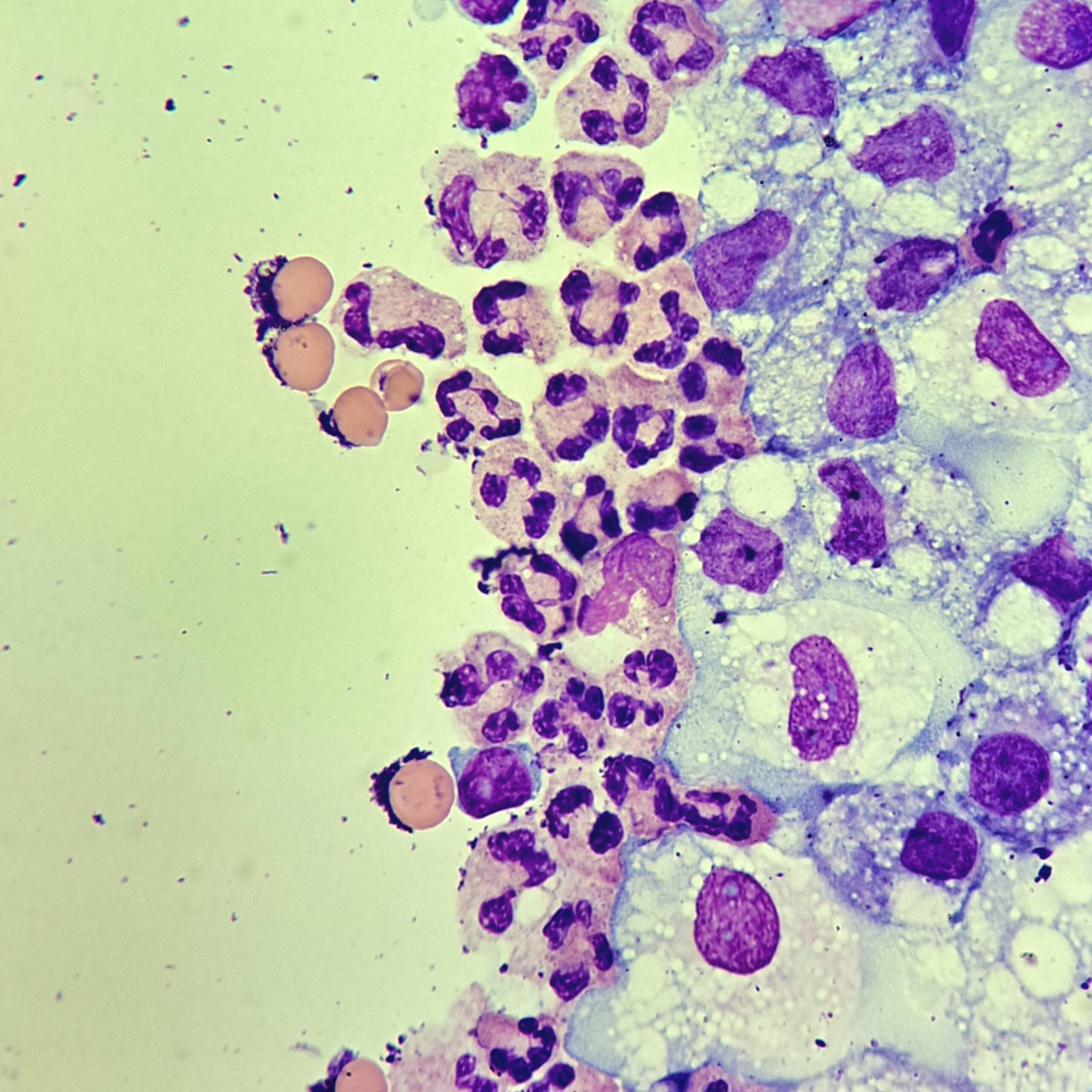
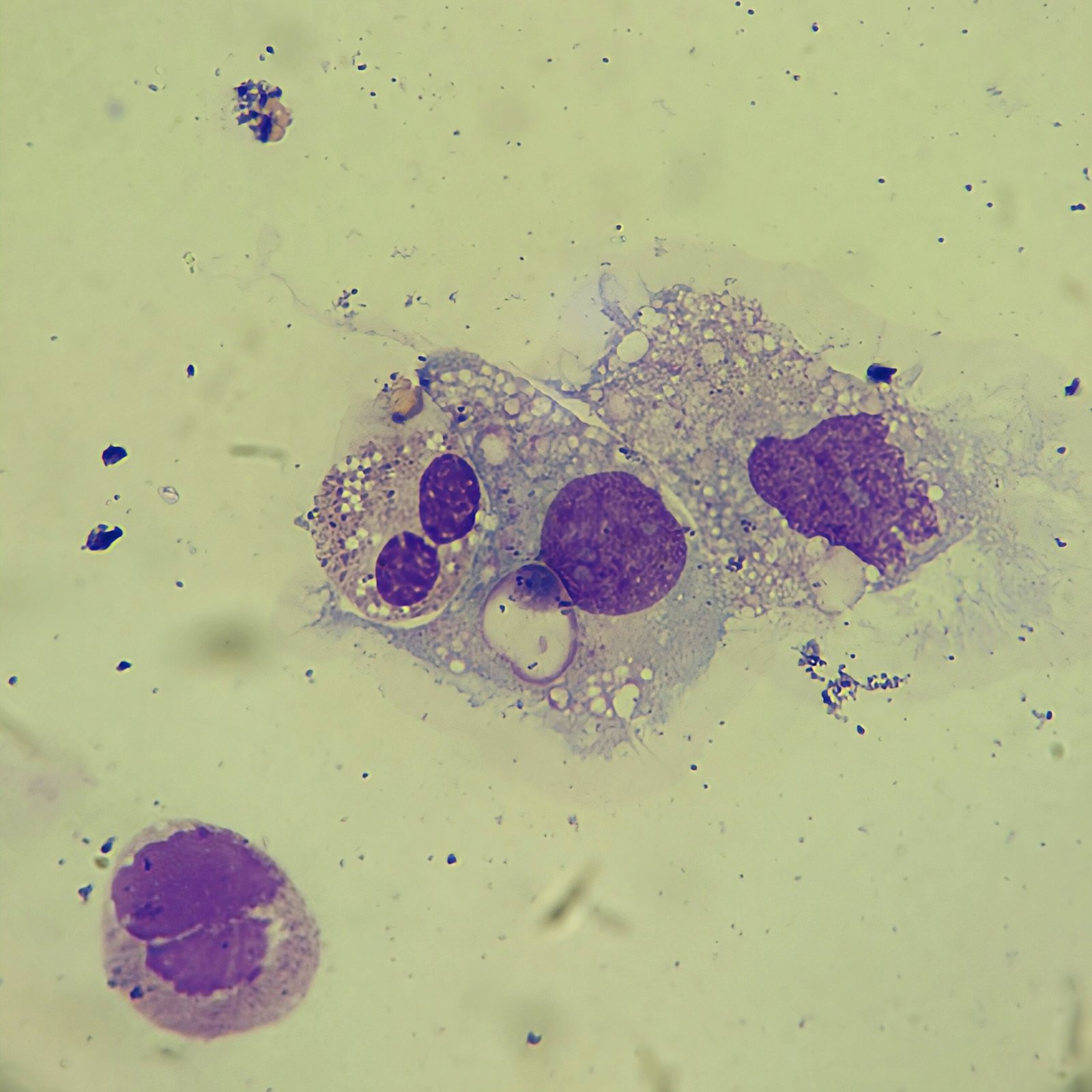
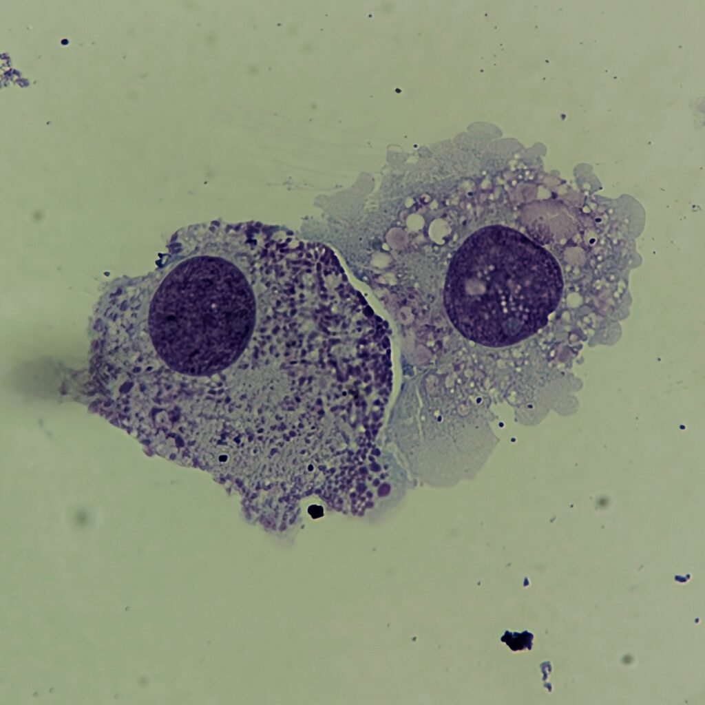
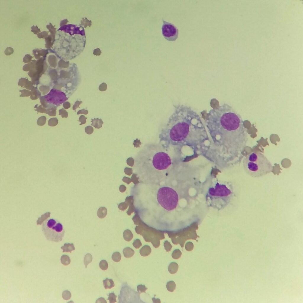
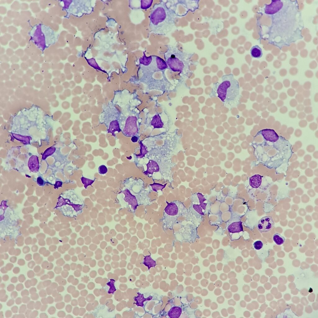
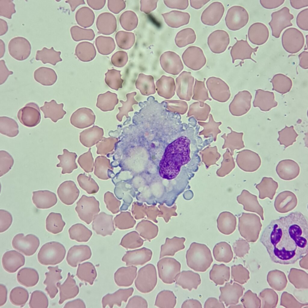
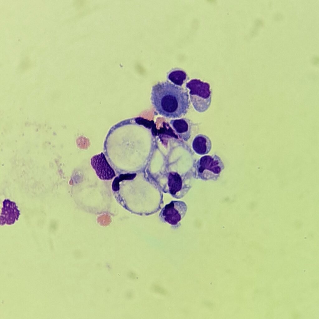
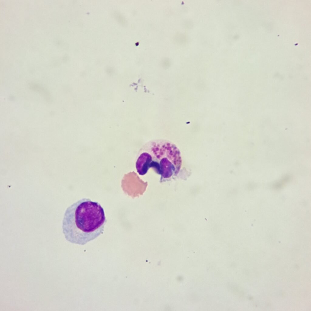
Monocytes / Macrophages
May appear as typical monocyte or as macrophage with vacuoles.
More Images
See More
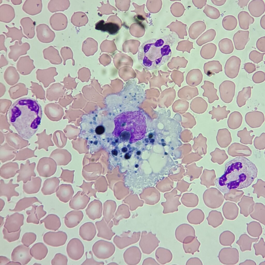
Erythrophage
Macrophage with engulfed RBCs.
Can be seen after hemorrhage. May also just be the result of the macrophage continuing phagocytic in vitro activity after collection.
More Images
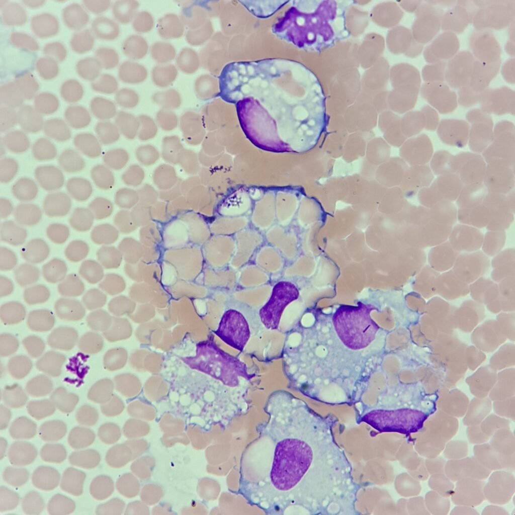
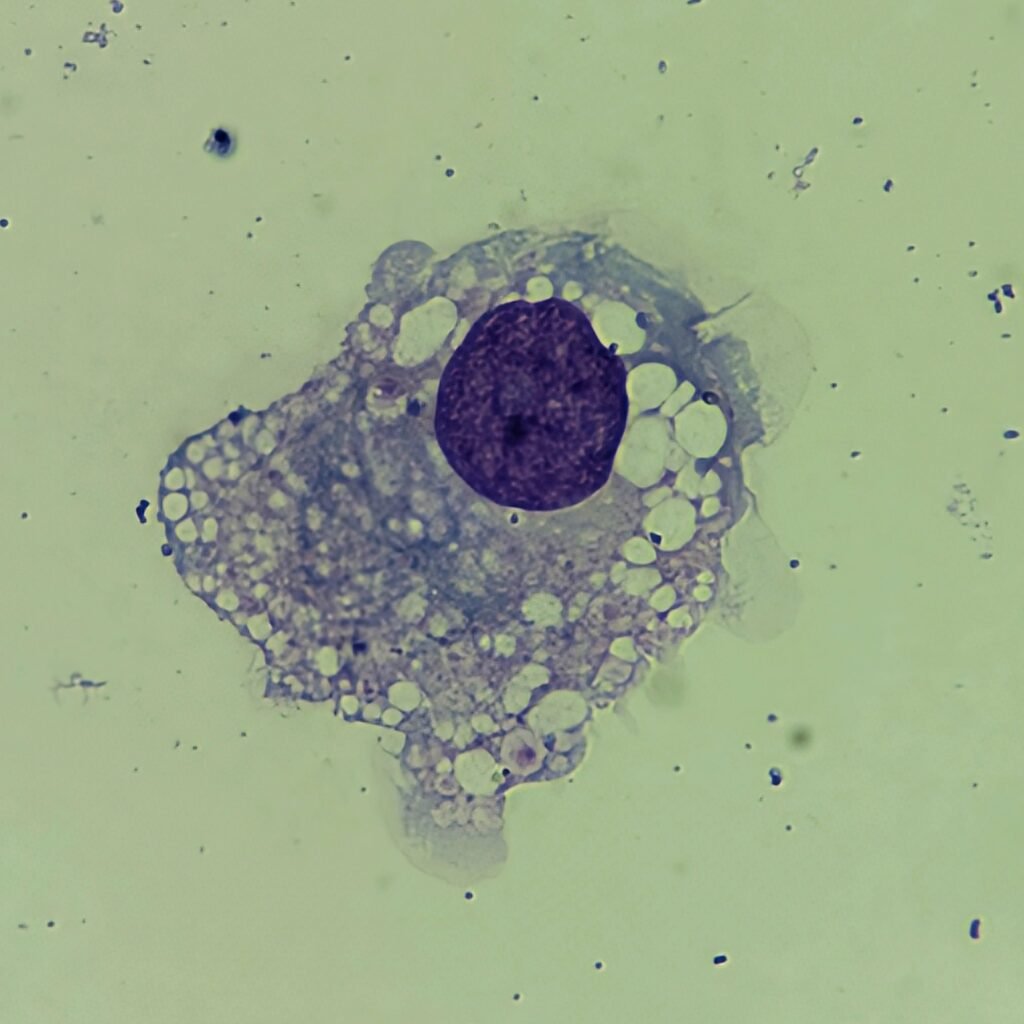
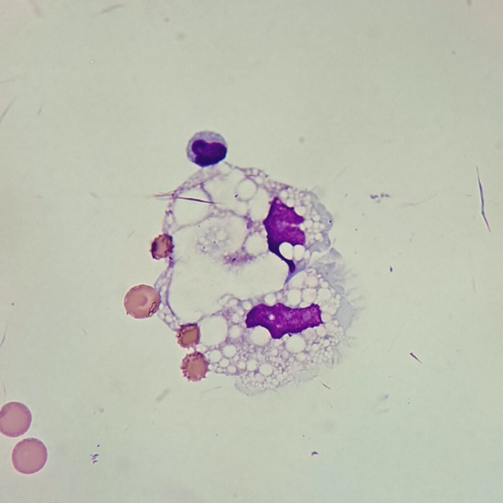
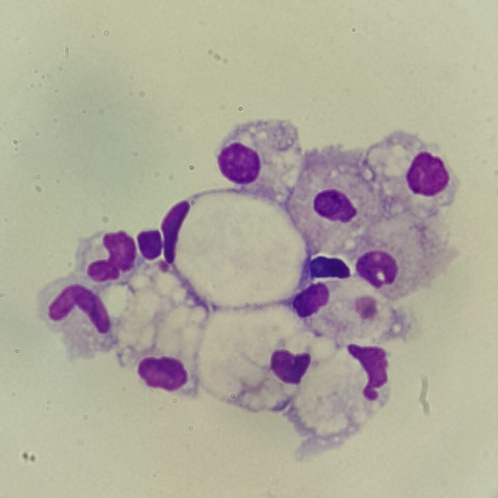
Lipophage
Macrophage with abundant lipid vacuoles. May appear in CSF following a brain infarct.
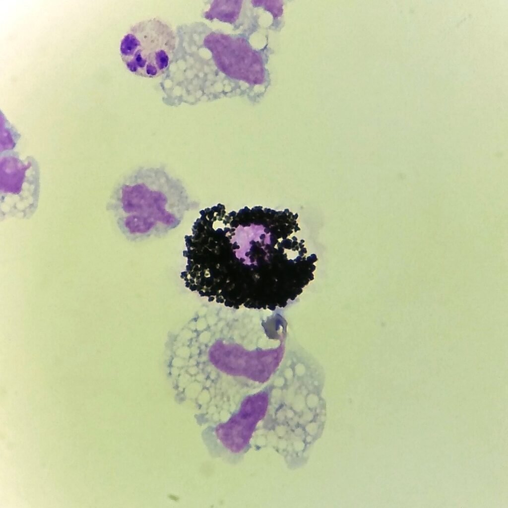
Siderophage
Macrophage with coarse dark blue granules .
Can be seen even months after hemorrhage as macrophages phagocytose the hemoglobin remains of RBCs.
May also be seen in disorders where there is excessive iron buildup.
More Images
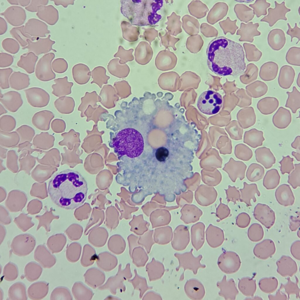
Signet Ring-Like
Macrophage has the appearance of a “signet ring” with several vacuoles forming what looks like the finger hole of a ring. The nucleus is pushed to one side of the cell, giving its distinctive appearance. Note that this type of macrophage is different from a true signet ring cell.
More Images
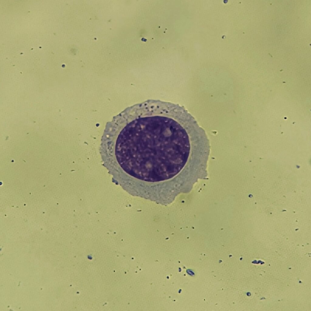
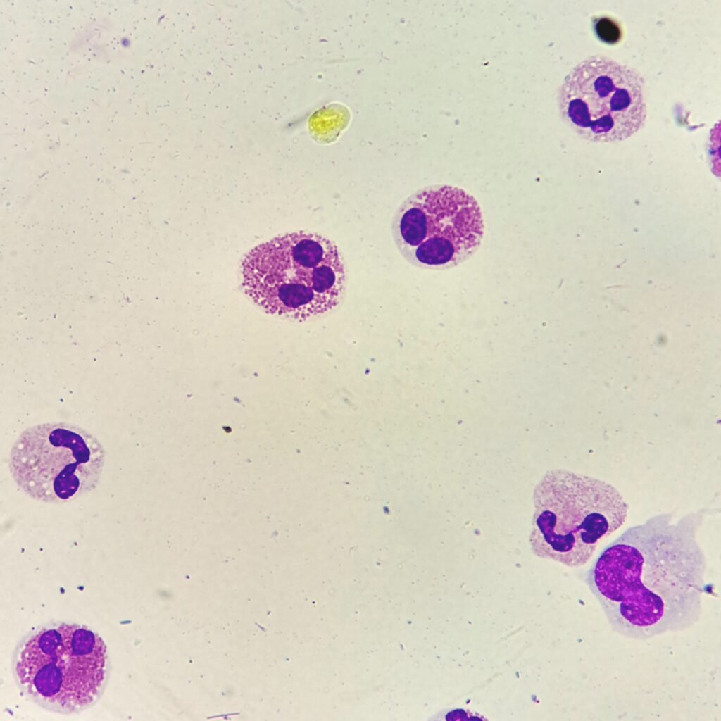
Eosinophils
Cells have coarse refractile reddish-orange granules.
Increased amounts may be seen with parasitic infection or foreign body reactions.
More Images
Basophils
Increased amounts may be seen with foreign body or allergic reactions.