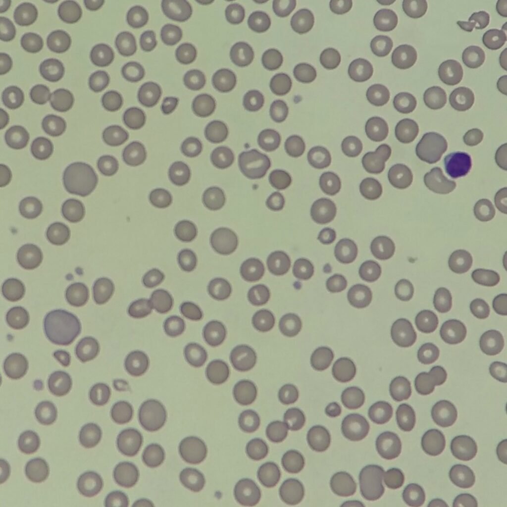
Auto Agglutination
RBCs clump together. This can cause erroneous results such as increased MCHC and MCV with a falsely decreased hematocrit.
Warm specimen at 37 oC to resolve.
Coming Soon 😀
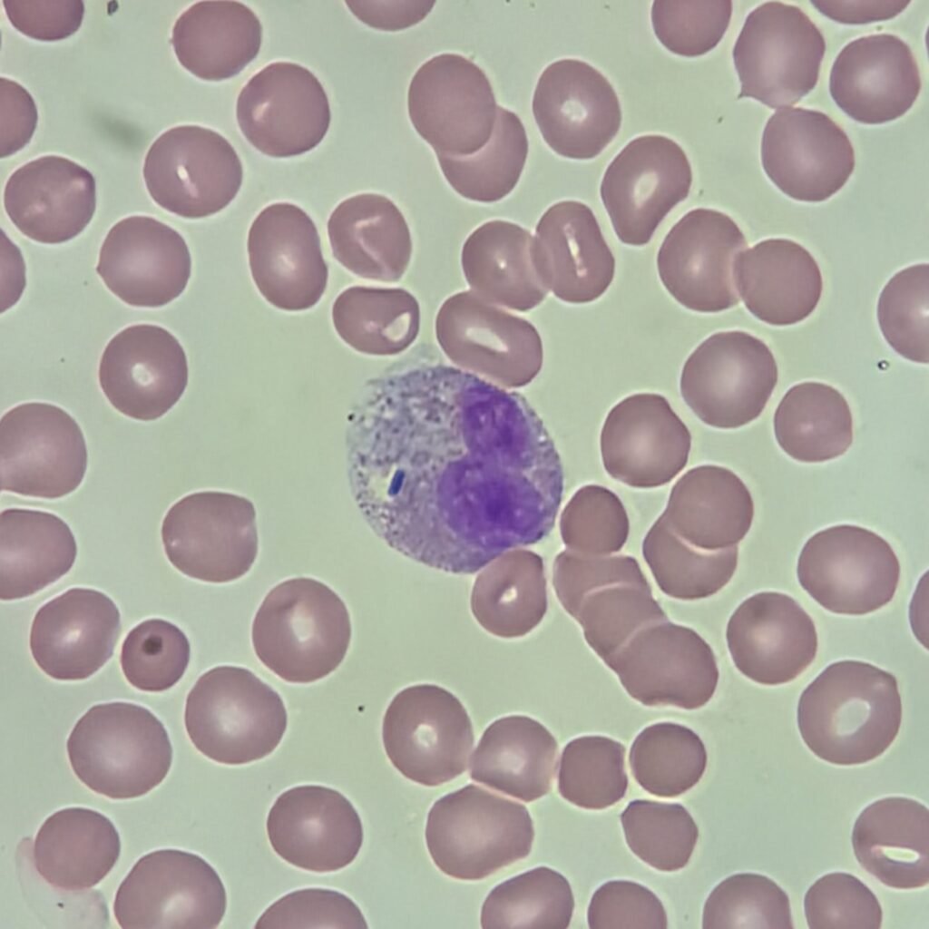
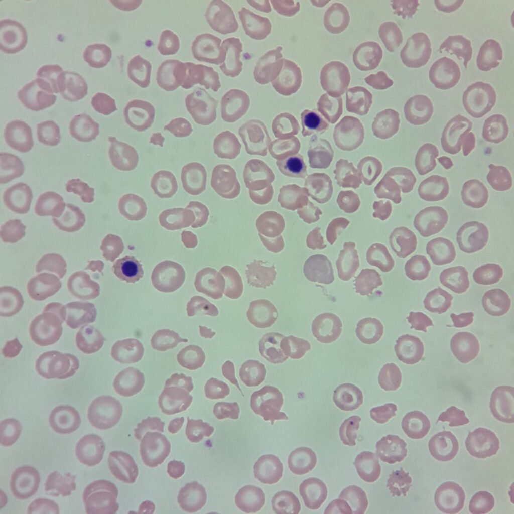
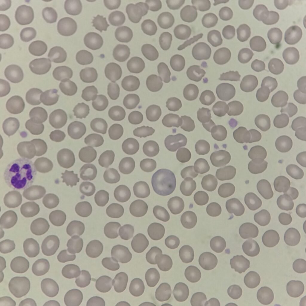
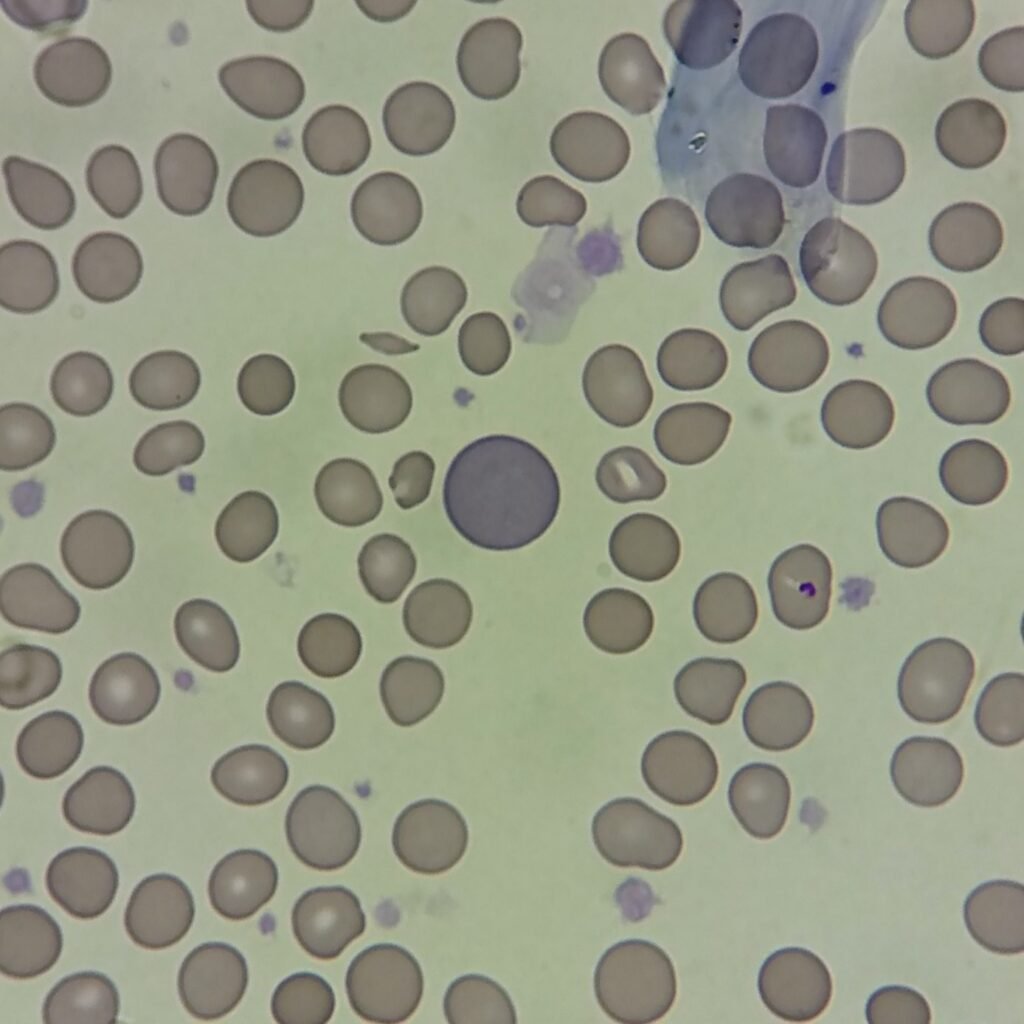
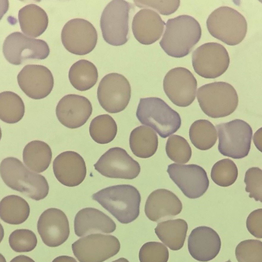
Polychromasia
RBC stains diffusely blue/gray due to residual RNA.
Slightly larger than mature RBC.
Proportional to reticulocyte percent but cannot be identified without supravital stain.
Seen in conditions with increased erythropoiesis such as hemolytic anemias or hemorrhages.
More Images
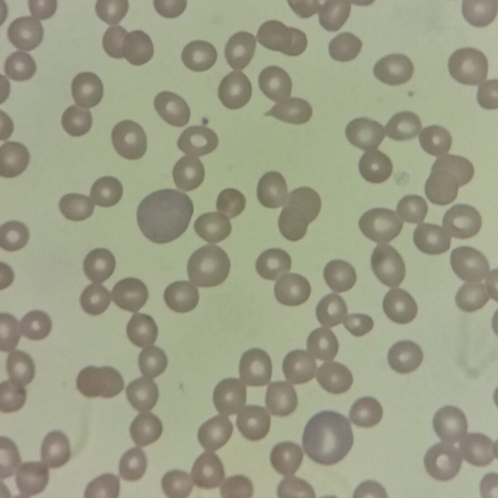
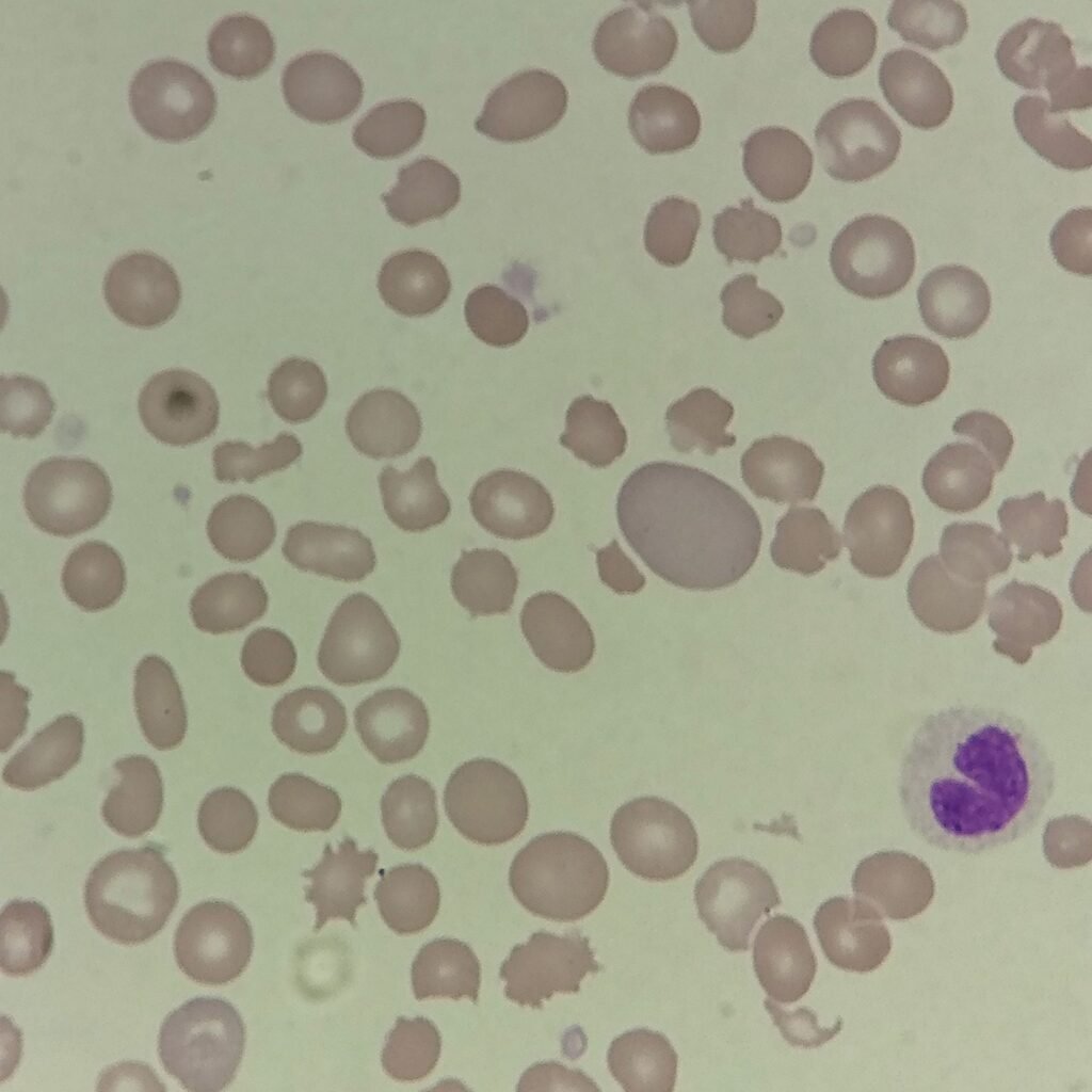
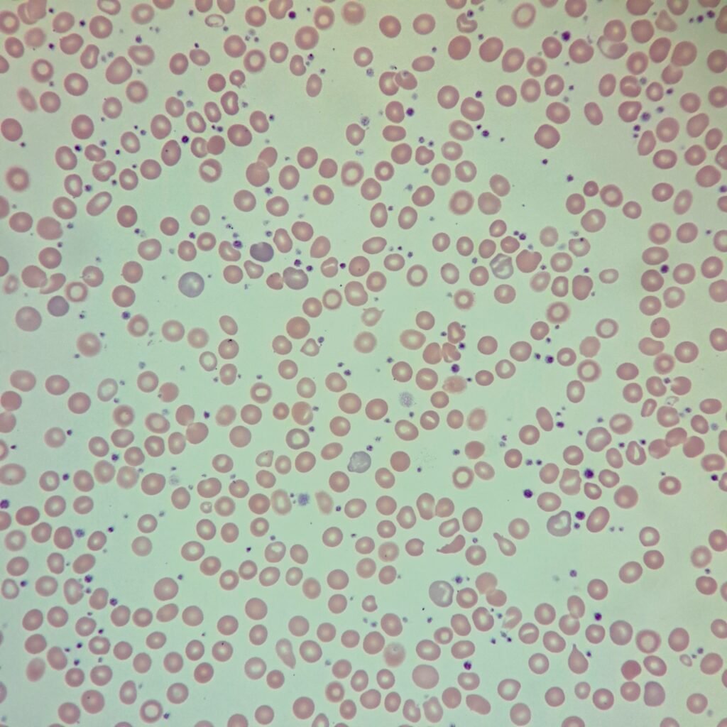
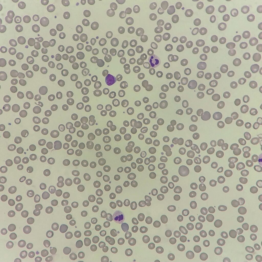

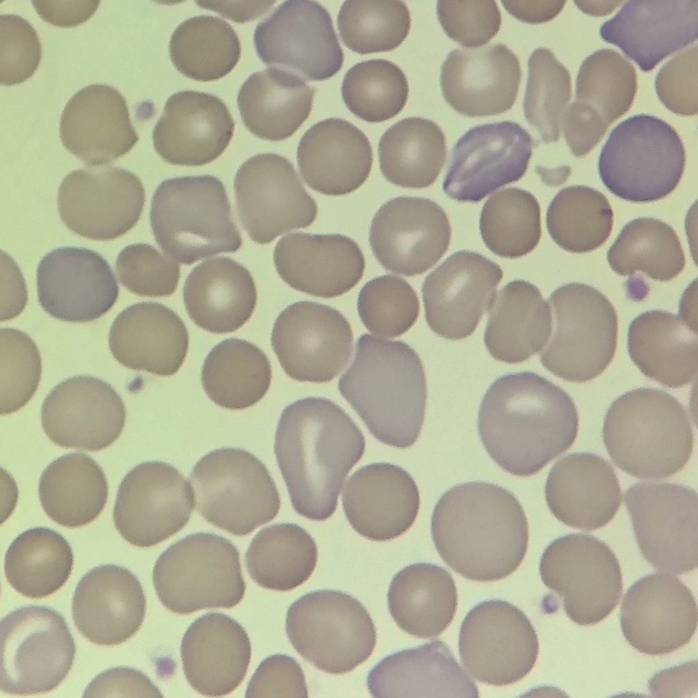
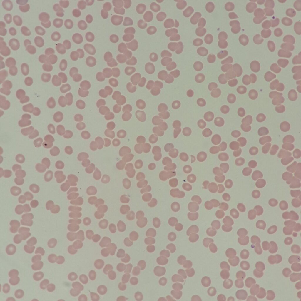
Rouleaux
RBCs stack together like coins in a linear fashion. Usually associated with increased plasma proteins, as seen in conditions like Multiple Myeloma. Can also be seen normally in thicker areas of the slide.
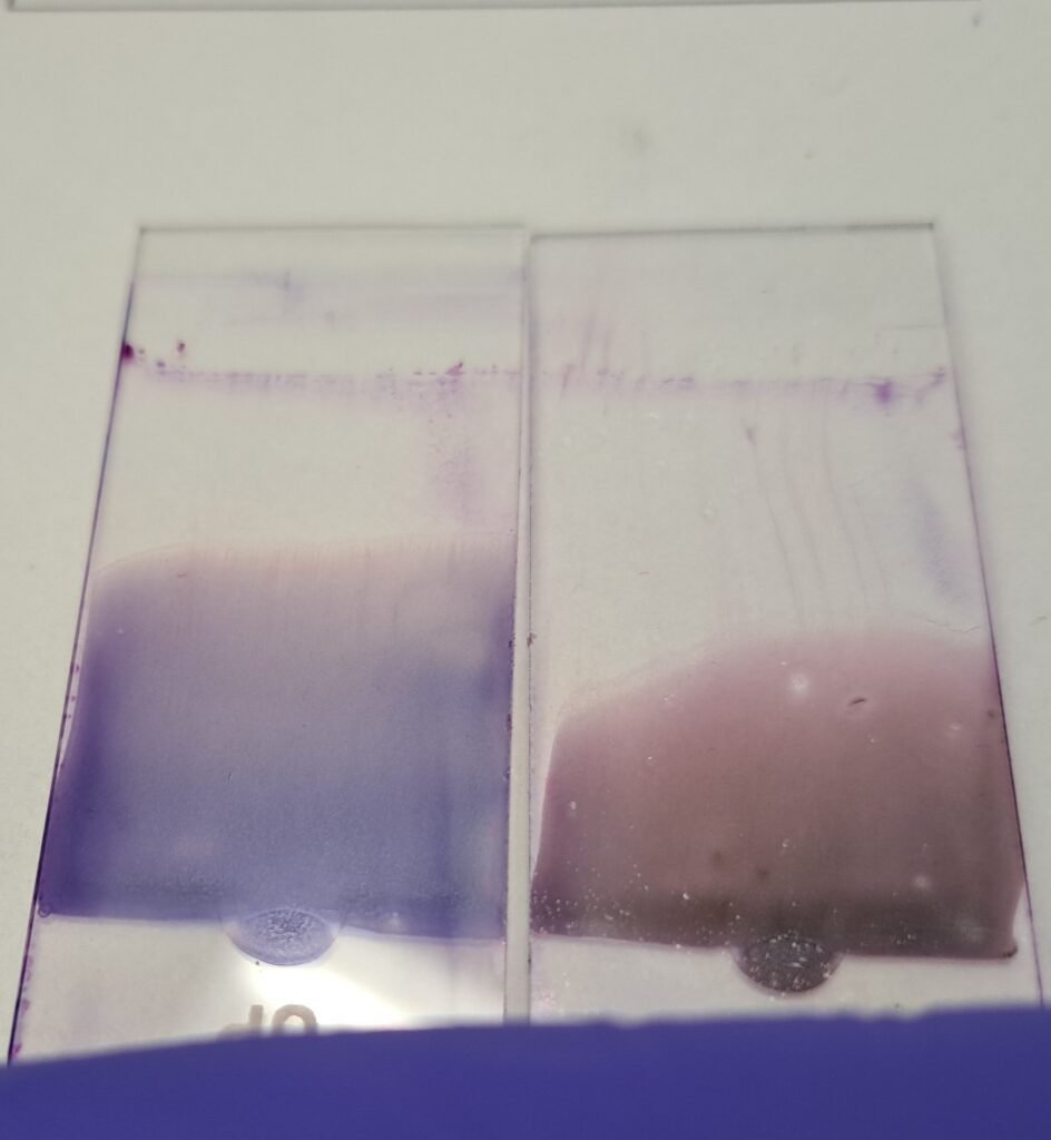
Excess proteins in blood may also cause the slide to stain more blue.
More Images