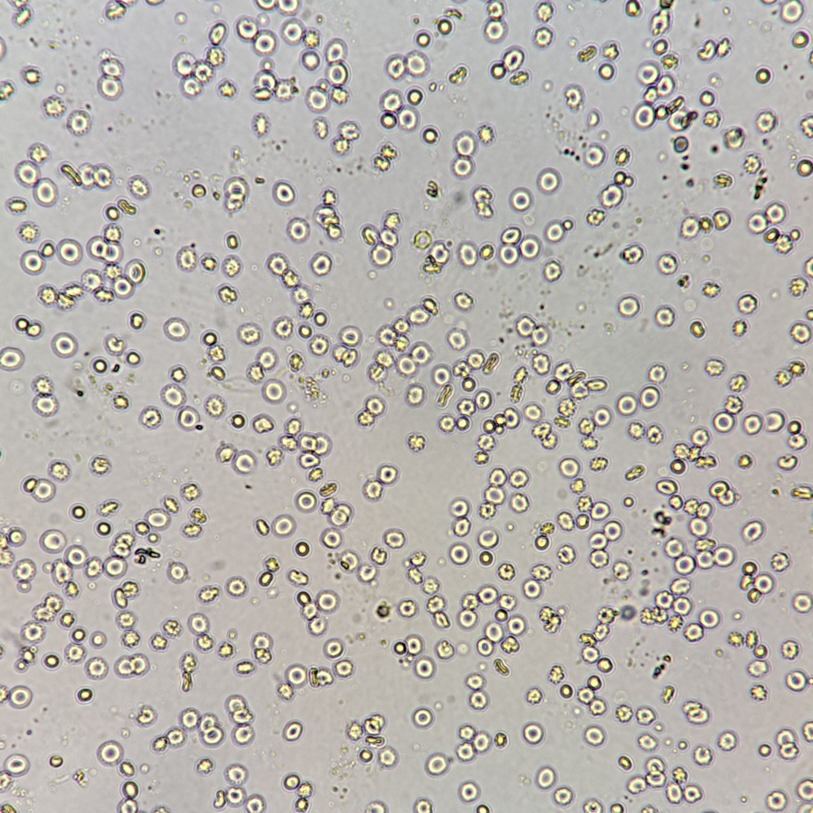
Urinary Casts
Casts are formed in the tubules of the kidney. Urinary stasis, lower pH, and increased solutes or proteins are all factors that can lead to cast formation. Usually accompanied by proteinuria.
Casts look like long cylinders, with parallel sides and round or blunted ends. Depending on where they were formed in the tubules, they may be straight or curved. It is important to distinguish casts from fibers.
The presence of casts is usually indicative of a form of renal disease.
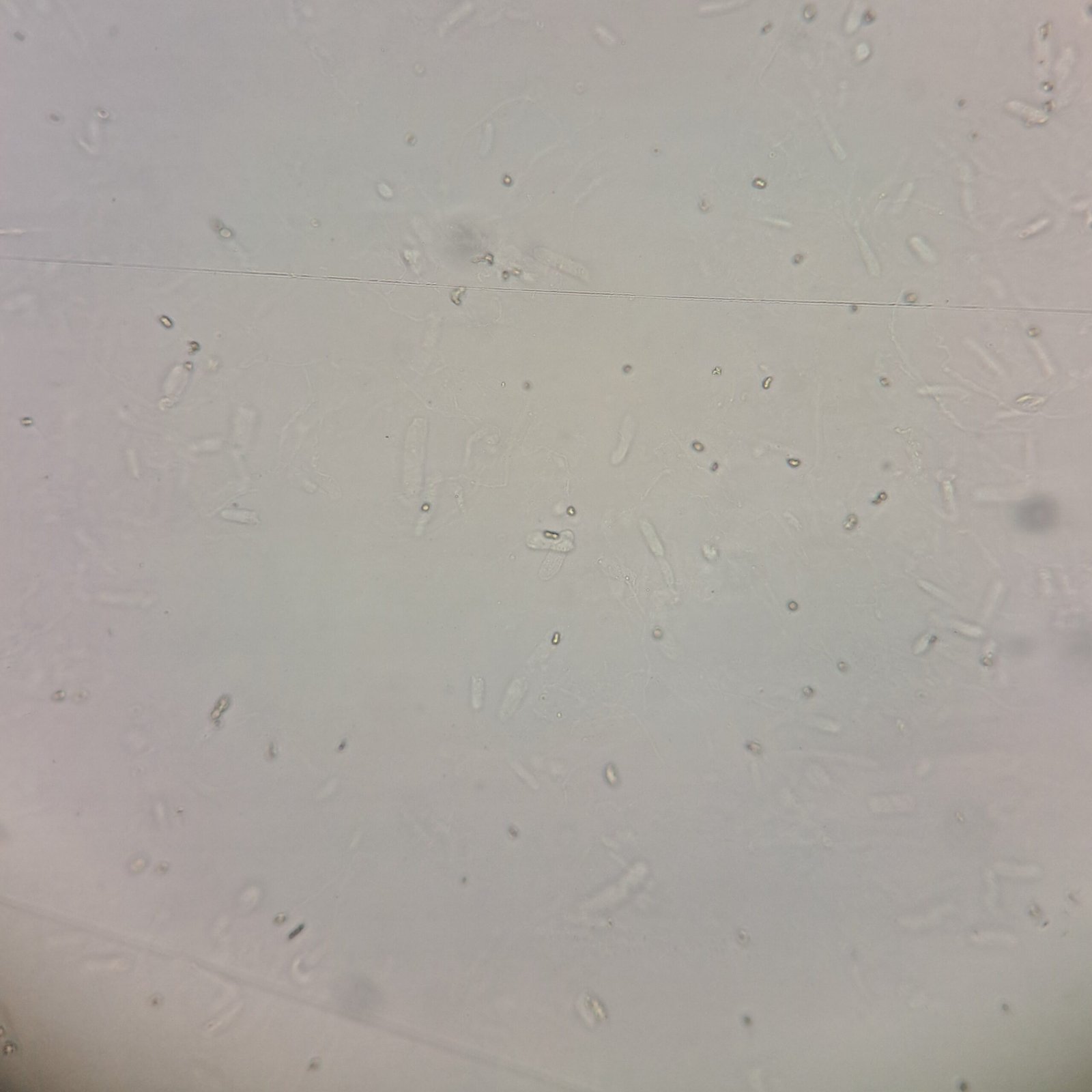
Hyaline Casts
Hyaline casts are translucent and have a low refractive index, making them difficult to see. Phase microscopy can be used to help with this.
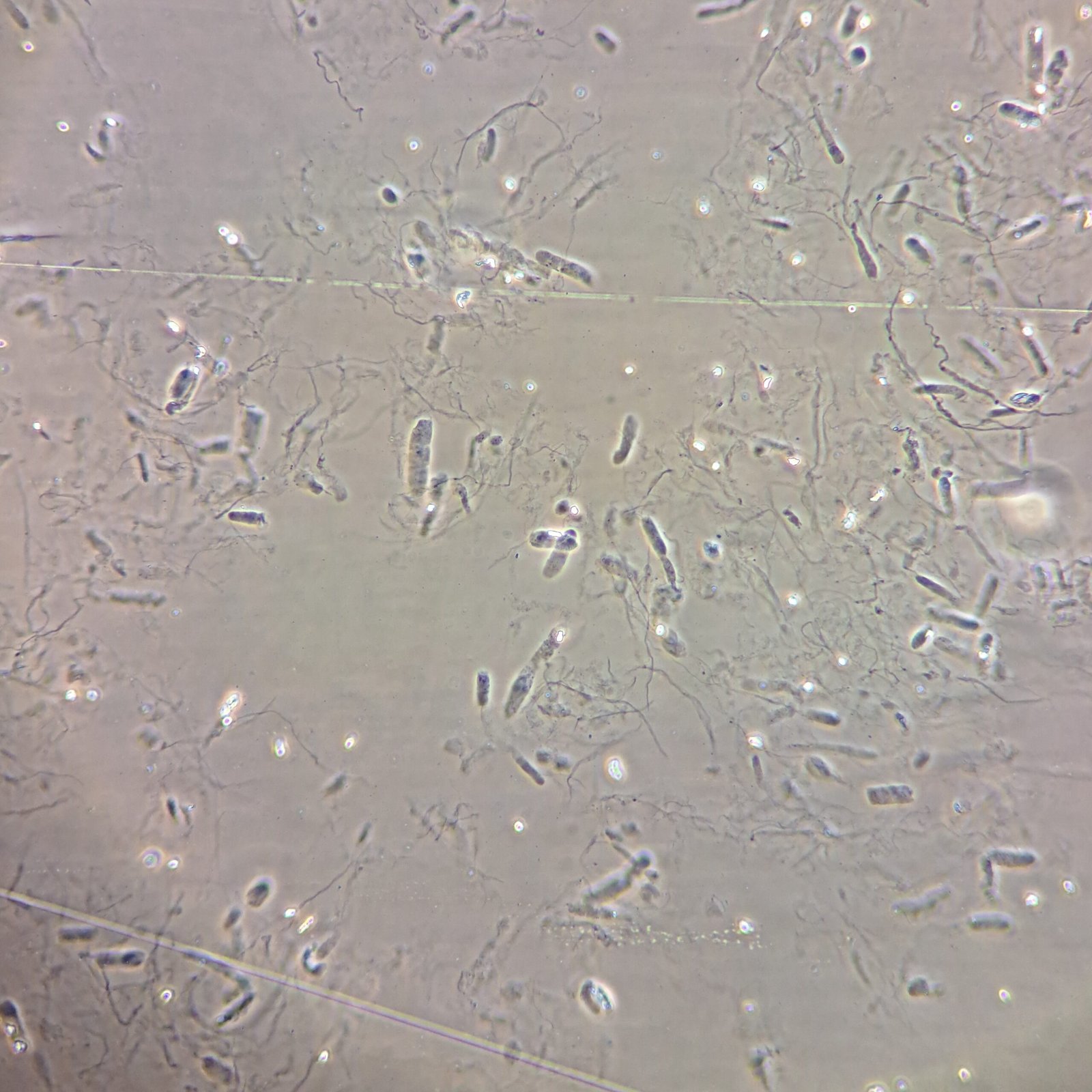
Granular Casts
Granular casts may contain fine or coarse granules. They should have a clearly defined shape.
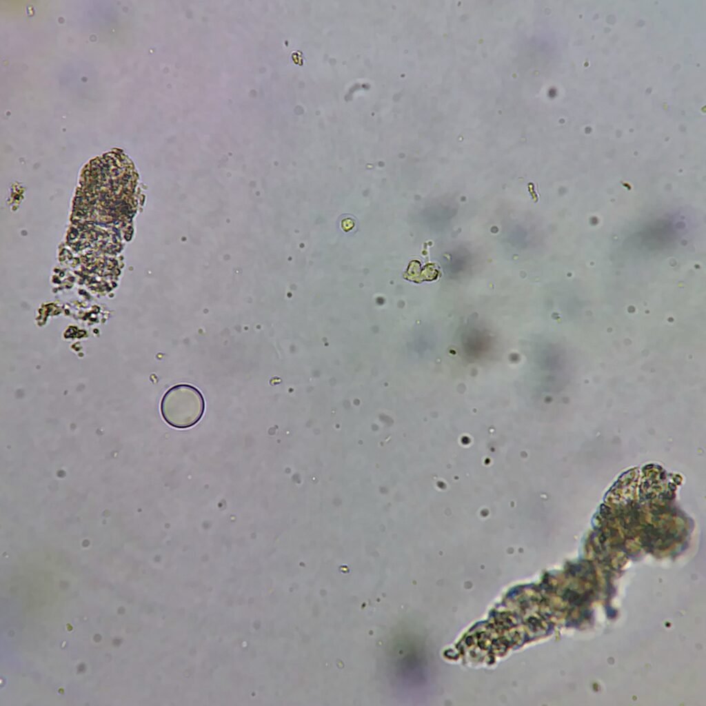
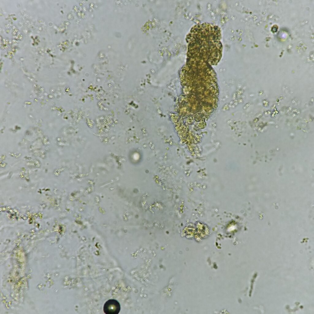
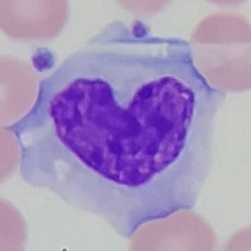
Red Blood Cell Cast
RBCs can be seen in a protein matrix. Many RBCs may color the cast yellow or reddish-brown.
RBC casts indicate renal hematuria. Diagnostic for acute glomerulonephritis but may be seen in renal trauma, infarction, and malignant hypertension.
No Image Yet 🙁
RTE Cell Cast
RTE casts contain renal tubular epithelial cells in a protein matrix. If the cells are too degraded to properly identify as RTEs, the casts may simply be called cellular casts.

Waxy Cast
Waxy casts have a high refractive index, giving them their waxy look. While the cast is still cylindrical in shape, it is more broad and may have blunt ends or cracks.