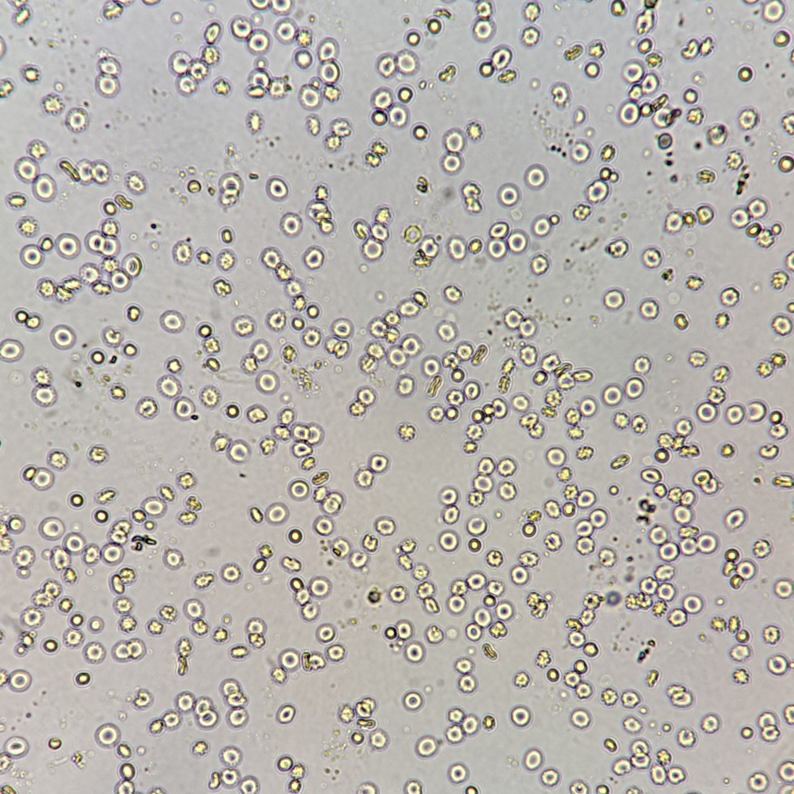
Red Blood Cells
RBCs are typically donut-shaped. However, they may also be crenated in hypertonic solutions or appear as an hourglass when viewed from the side. RBCs can be identified by their thin dark borders and high refractive index. Dysmorphic RBCs can be confused for budding yeast.
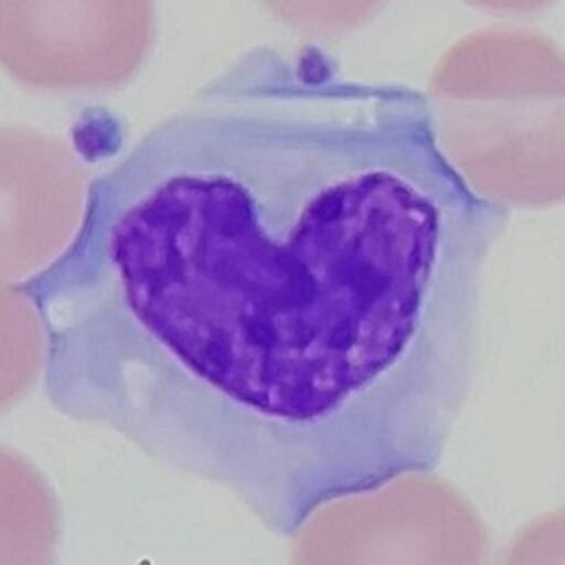
White Blood Cells
WBCs can vary in size depending on the cell. They are round with granular cytoplasm and a nucleus, unlike RBCs. WBCs may also appear in clumps.

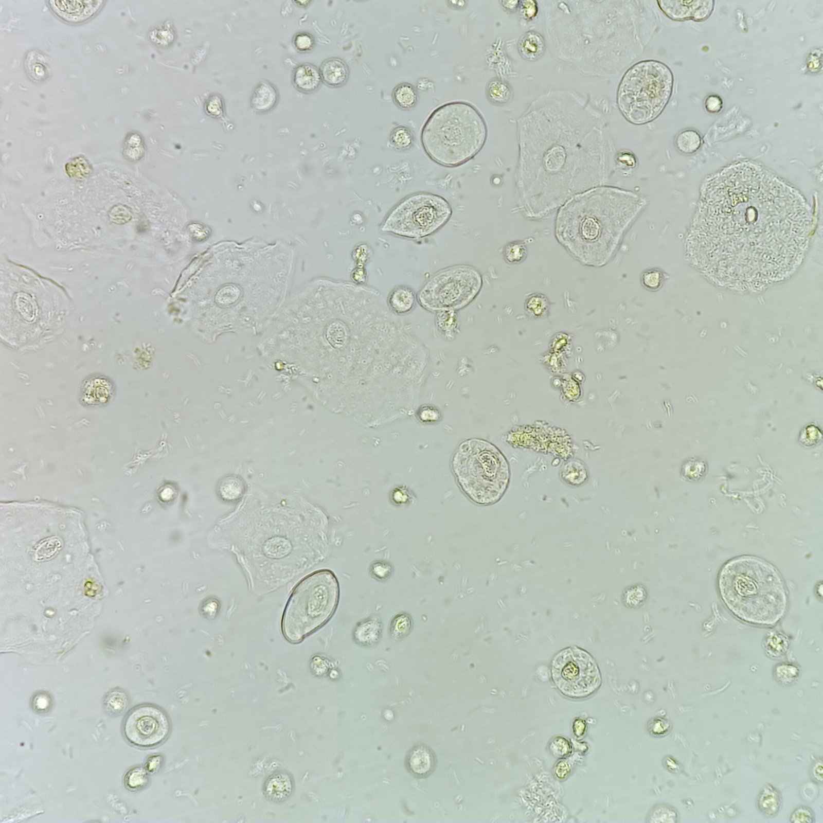
Squamous Epithelial
Squamous epithelial cells are large with abundant cytoplasm and a small nucleus. Edges may be folded over or curled up.

Transitional Epithelial
Transitional cells are round or pear-shaped. They have less cytoplasm and a larger nucleus than squamous cells. Their defined border and readiness to absorb water can give them a bubble-like appearance.
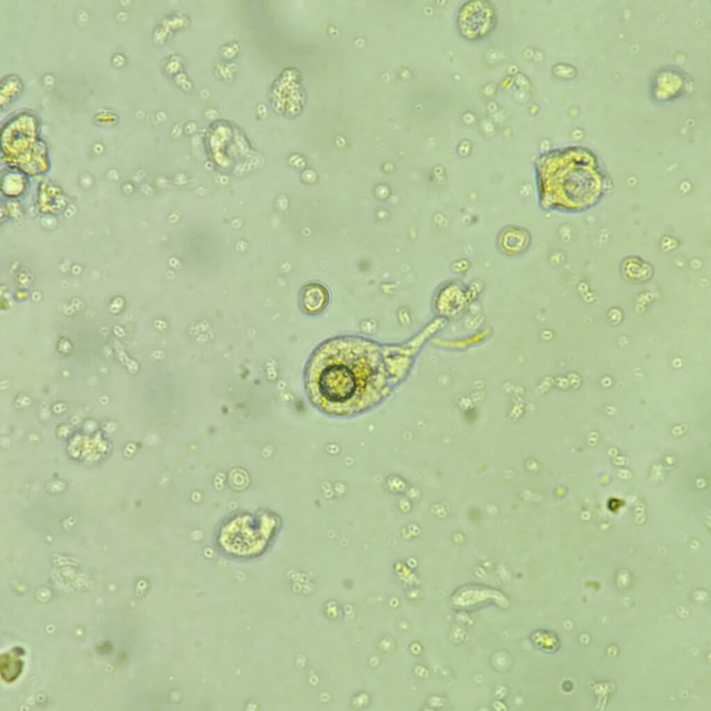
Renal Tubular Epithelial
RTEs are slightly larger than a WBC and may be round, oval, or polyhedral. Cytoplasm is finely granular, and nucleus is distinctly round. Difficult to distinguish from transitional epithelial cells.
Increased amounts indicate tubular damage, such as acute tubular necrosis, viral infection, transplant rejection, or drug or heavy metal toxicity.
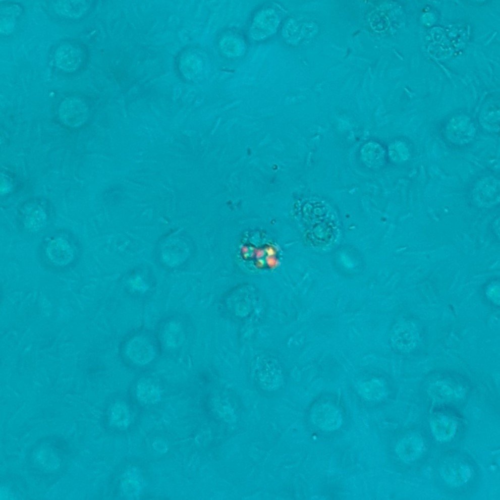
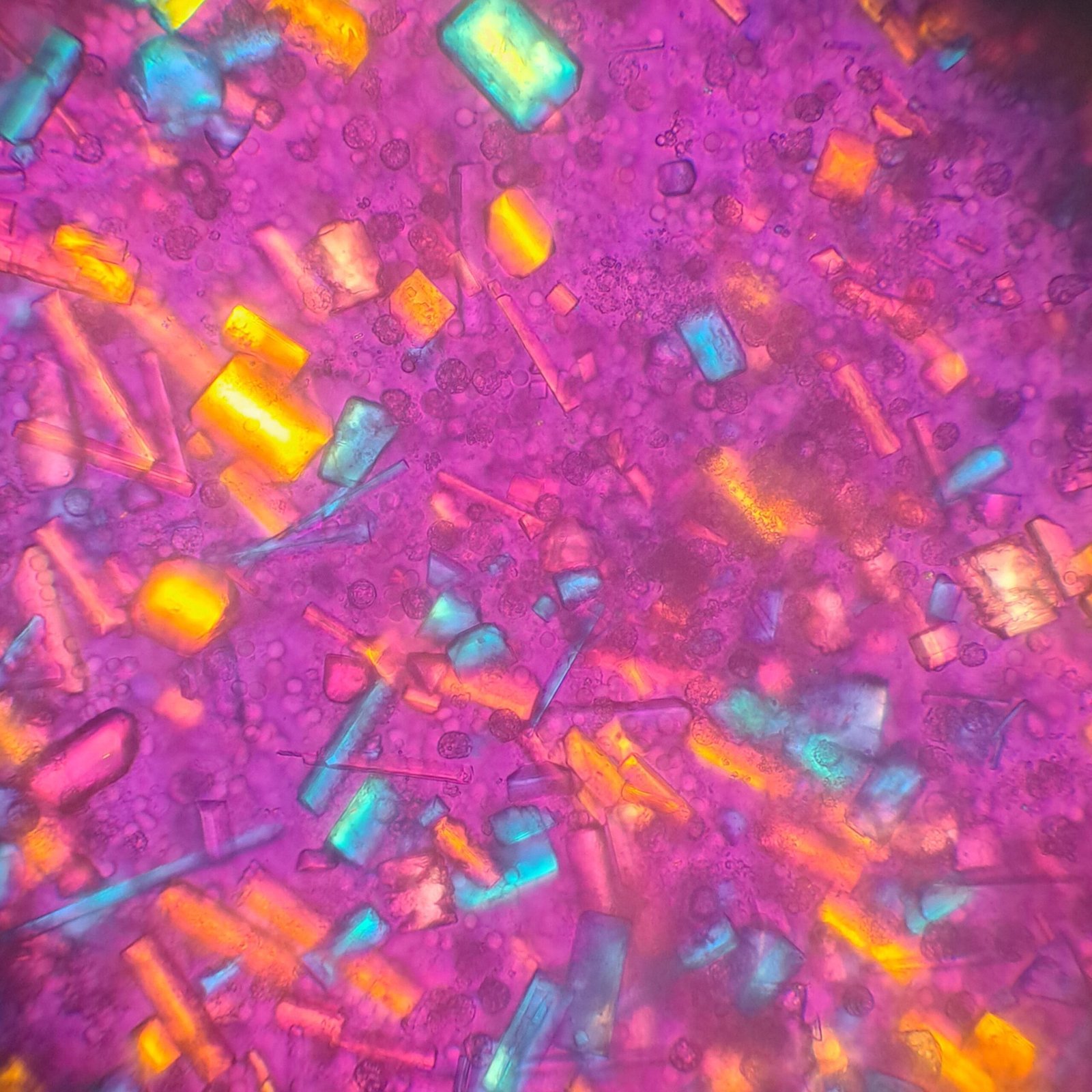
Oval Fat Bodies
Oval fat bodies usually renal tubule epithelial cells containing fat droplets, though they may also be leukocytes that have digested lipids. These fat droplets are highly refractile and form a “Maltese cross” pattern when polarized.