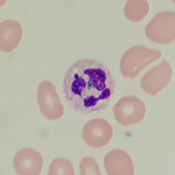
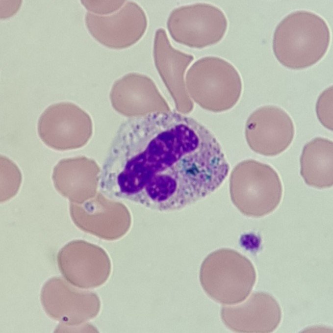
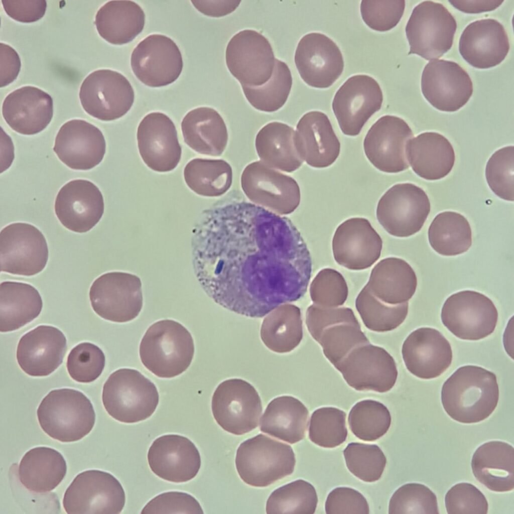
Dohle Bodies
Cytoplasm contains one or more blue/gray inclusions, usually along the periphery. May be accompanied with toxic granulation or vacuolization.
Seen most often in infection.
More Images
Toxic Granulation
Cytoplasm contains coarse, dark blue granules. May be accompanied with dohle bodies or vacuolization.
Seen most often in infection.
More Images


Vacuolization
Cytoplasm contains vacuoles, indicating phagocytosis. May be accompanied by dohle bodies or toxic granulation.
Seen most often in infection or inflammation. Can also be seen as a result of cell degradation.
More Images
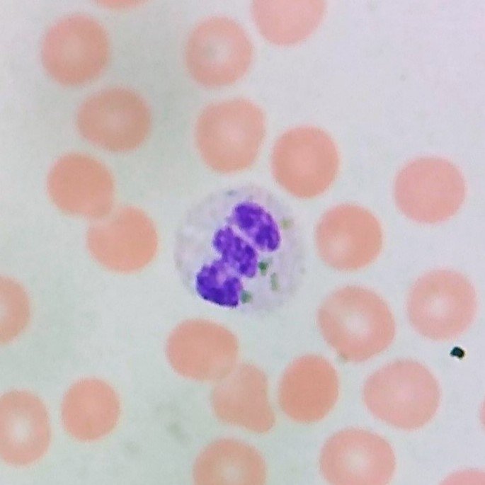
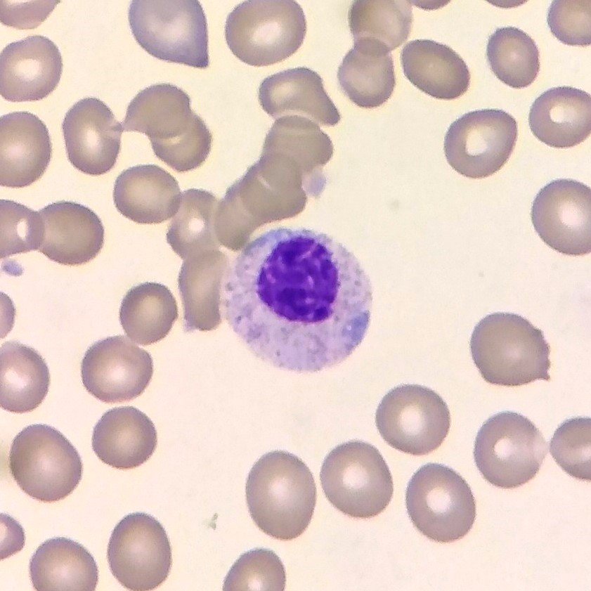
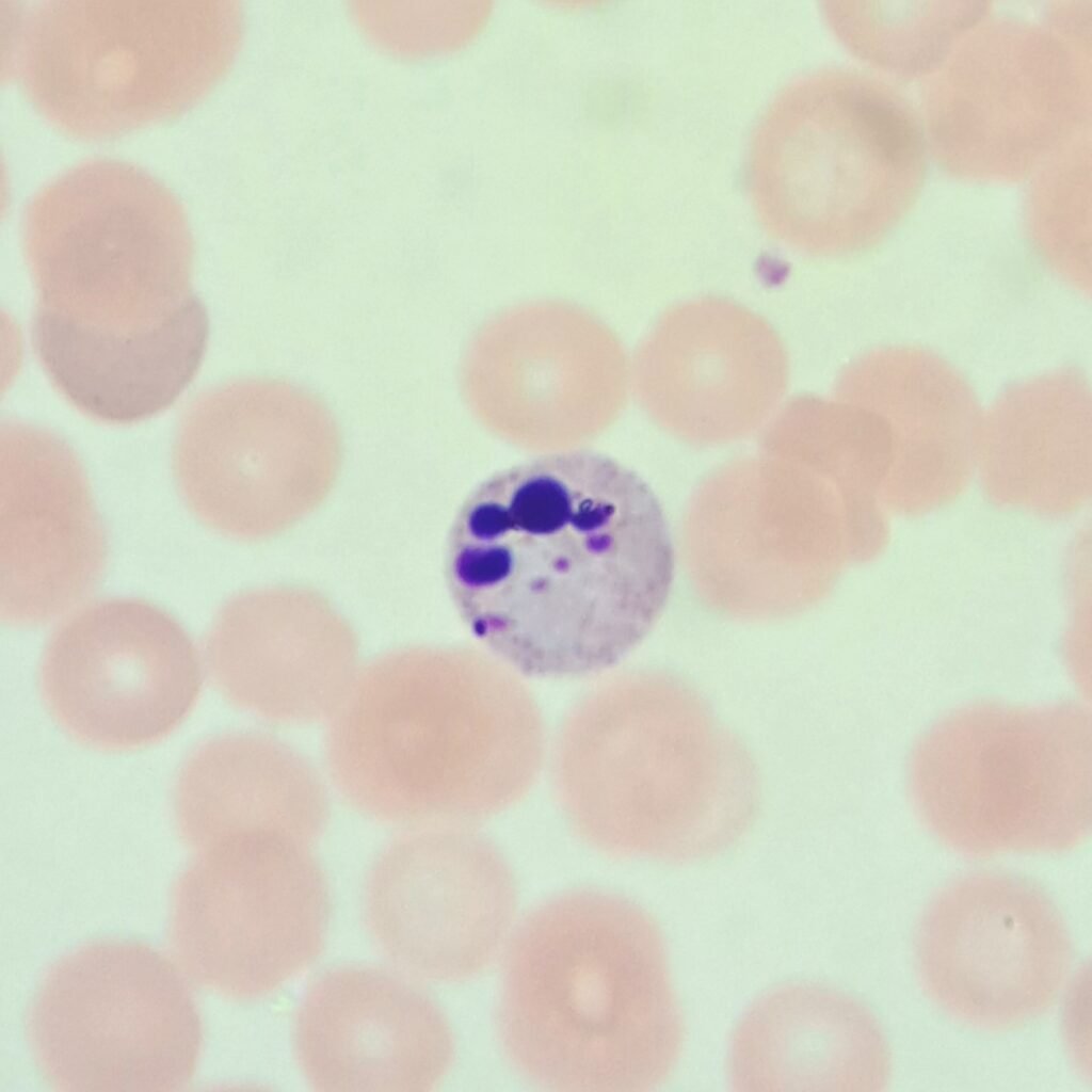
Critical Green Inclusions
AKA “Green Crystals of Death” due to being associated with poor prognosis.
Bright green/blue refractile inclusions in the cytoplasm.
Rare finding typically seen in critically ill patients.
More Images
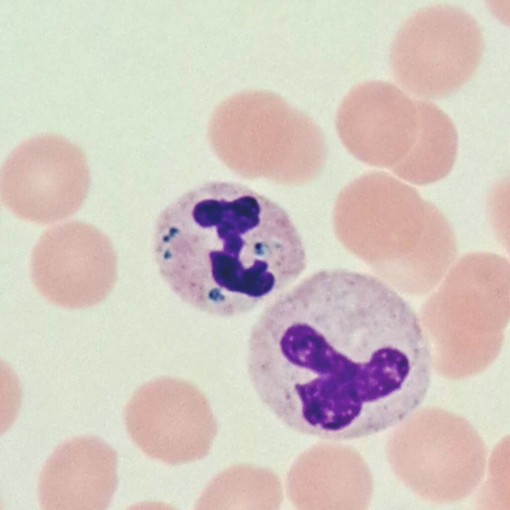
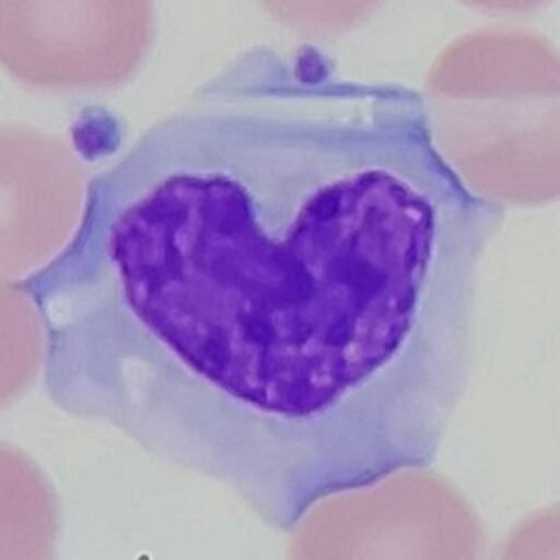
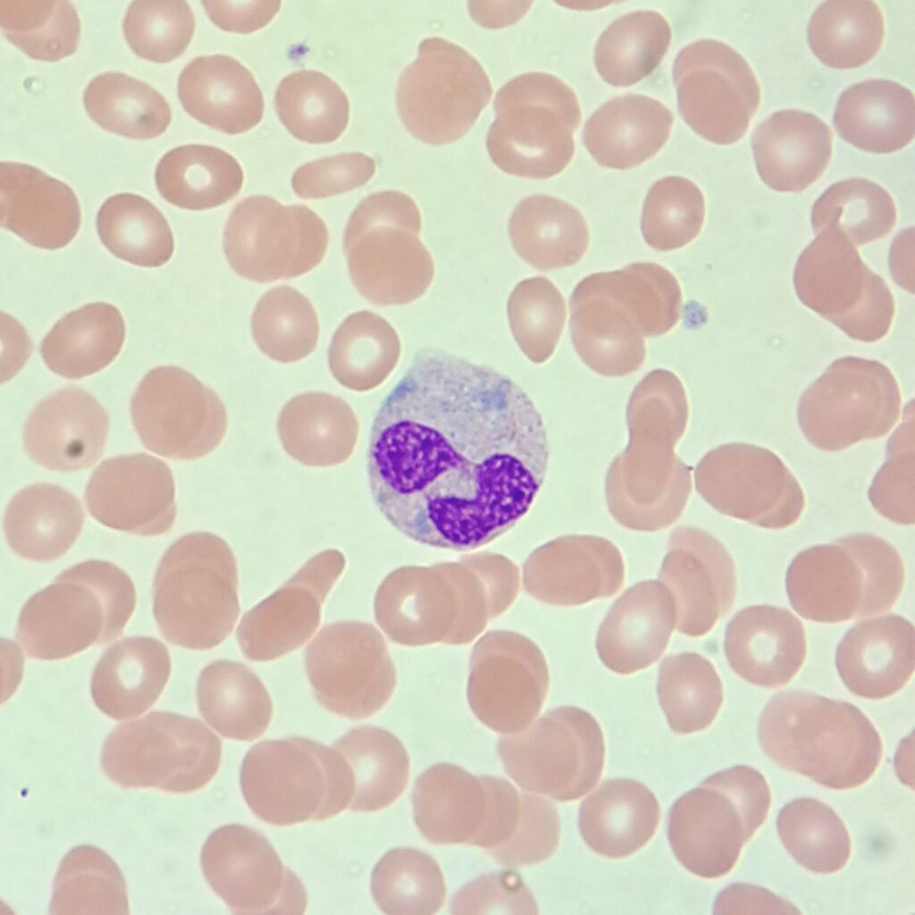
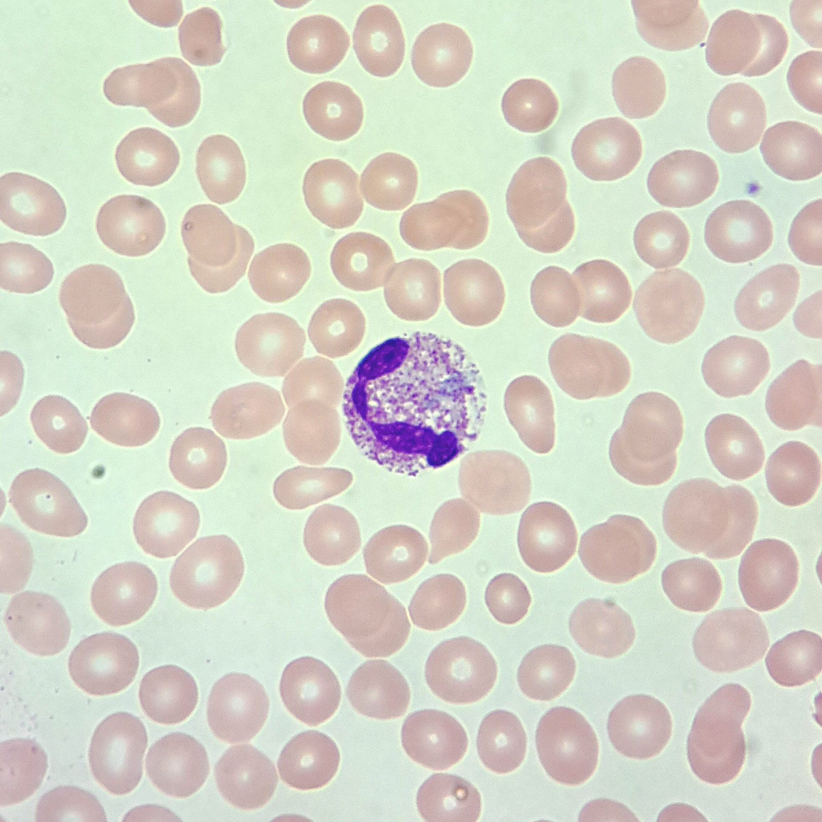
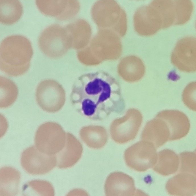
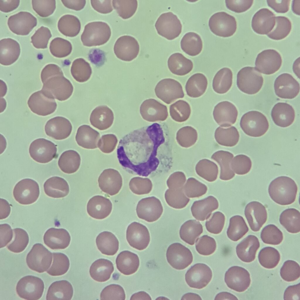
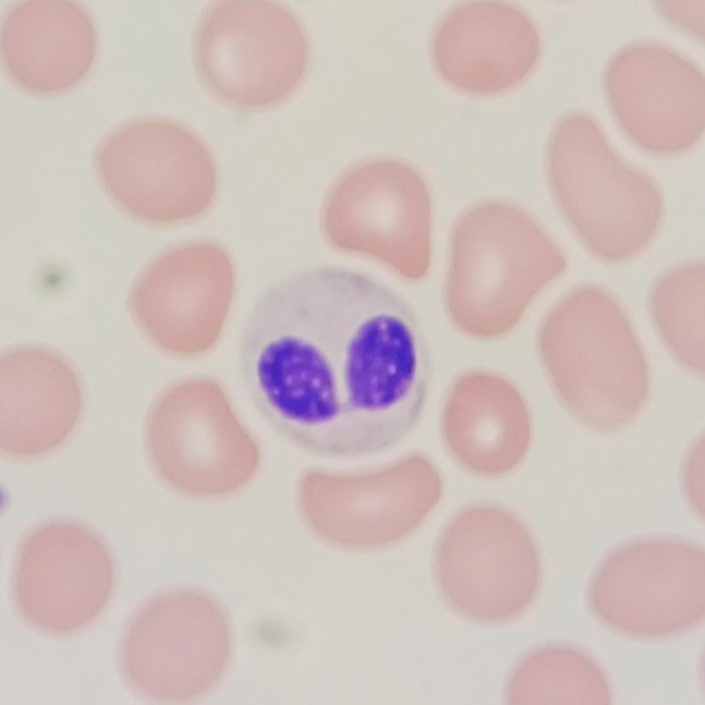
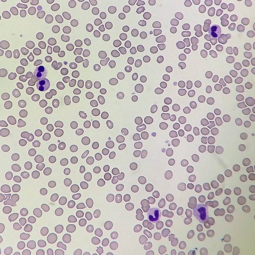
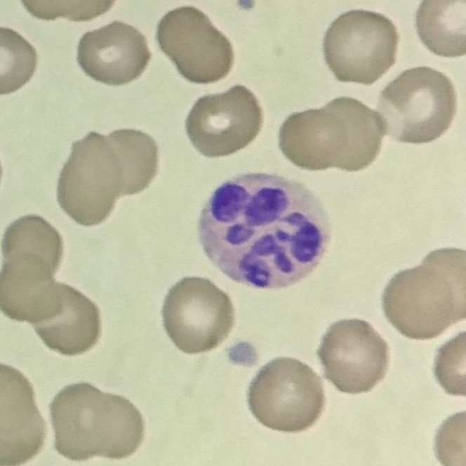
Pelger-Huet/Pelgeroid
Nucleus is hyposegmented, either bilobed or monolobed. Bilobed neutrophils are described as having a characteristic pince-nez shape (a particular style of glasses). Monolobed neutrophils may be mistaken as myelocytes if care is not taken. However, chromatin is notably condensed upon closer inspection
True Pelger-Huet neutrophils are an inherited disorder and completely benign. The majority of neutrophils will be hyposegmented.
Pseudo-Pelger-Huet, or pelgeroid cells, can be caused by certain medications or Myelodysplastic Syndromes.
More Images
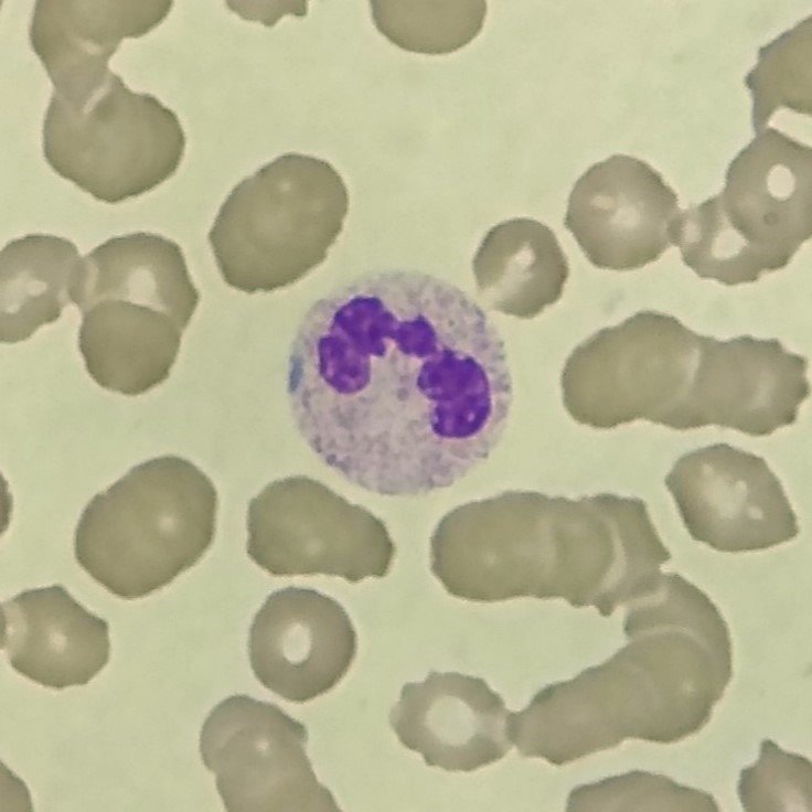
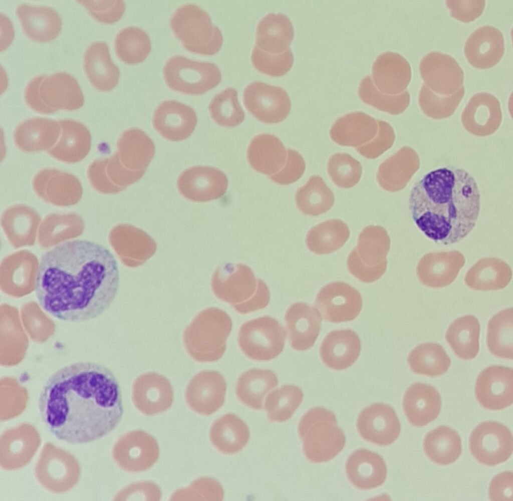
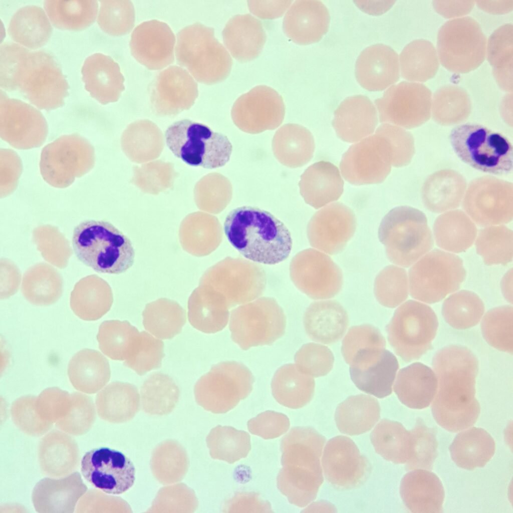
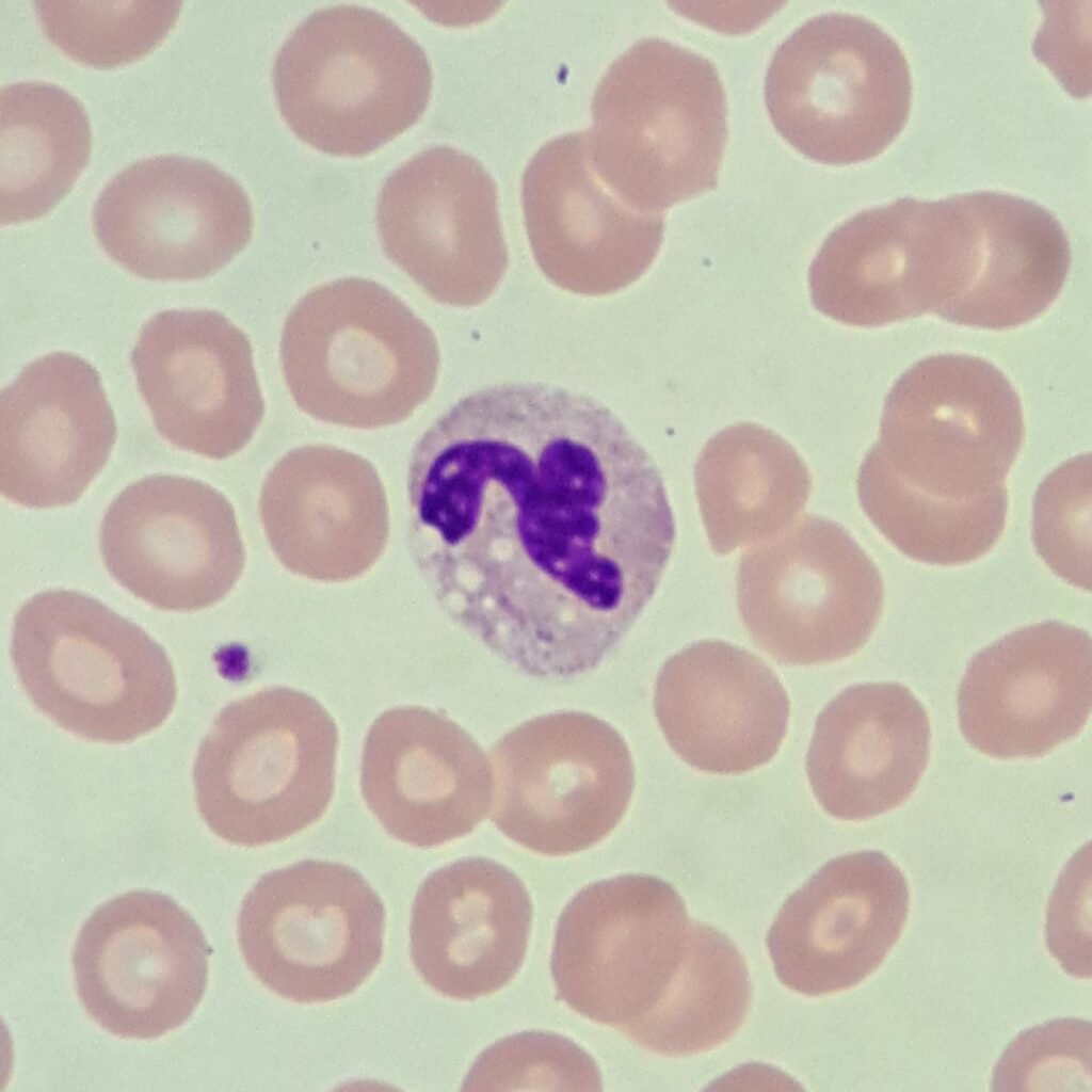
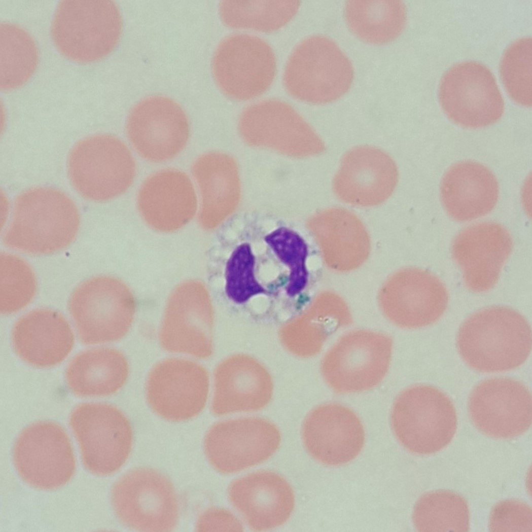
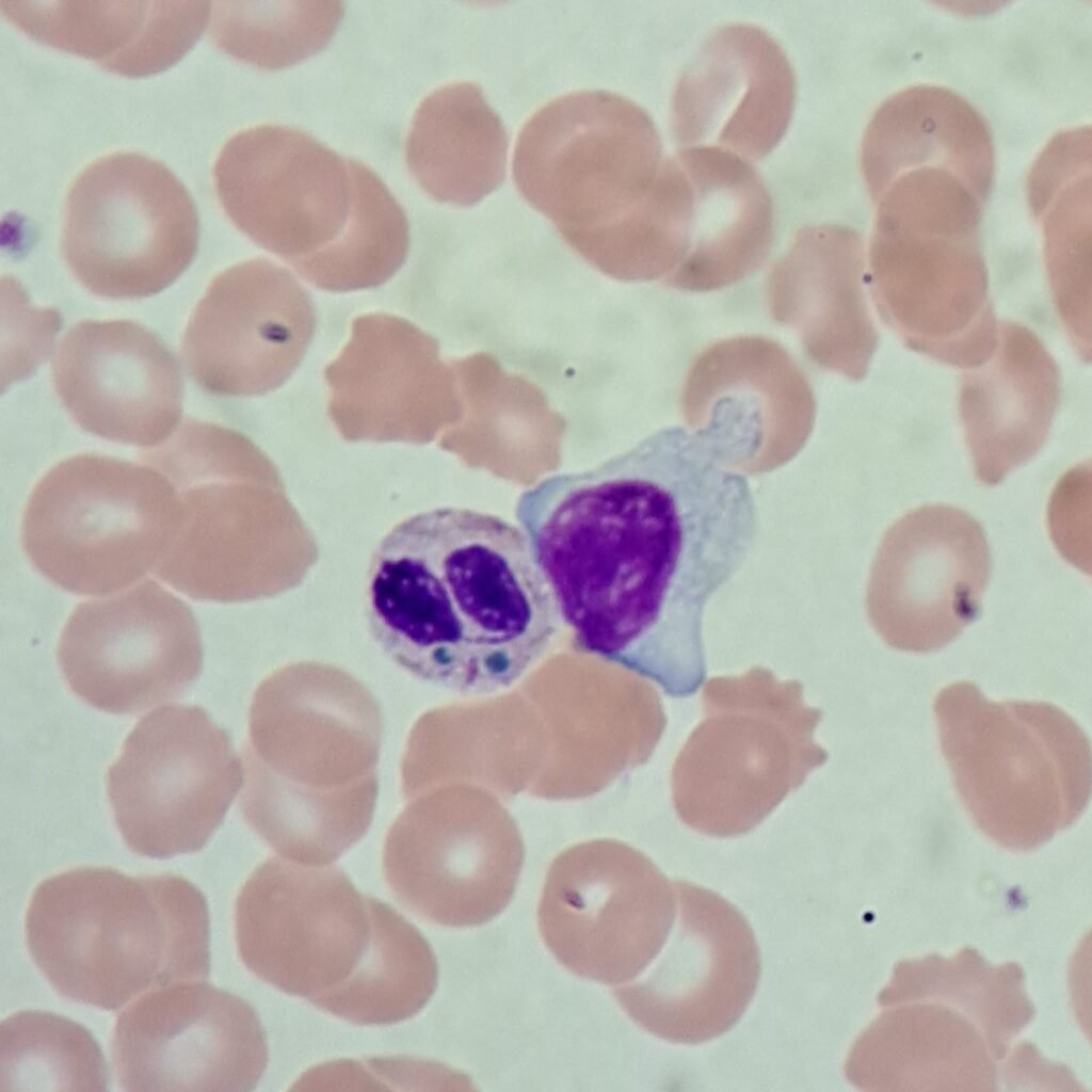
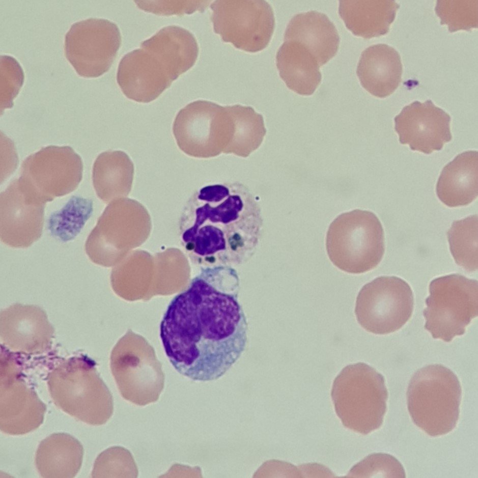
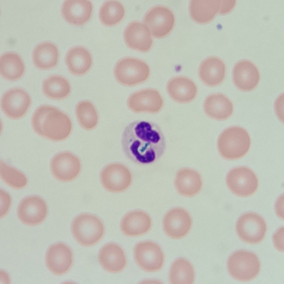
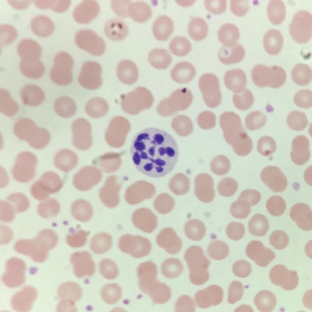
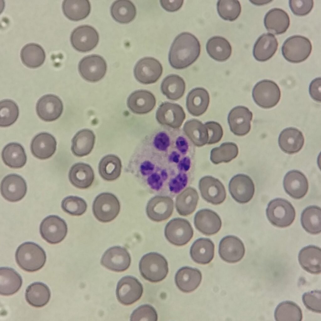
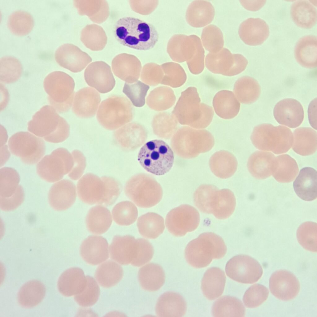
Hypersegmentation
Nucleus has 6 or more lobes. Cell is usually larger than normal neutrophils.
Can be hereditary. Also seen in Megaloblastic hematopoiesis due to nutritional deficiencies.
More Images
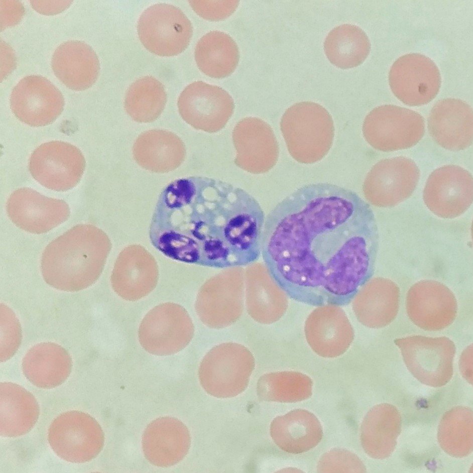
Pyknotic Neutrophil
Degenerated neutrophil. Nucleus stains densely blue or purple. Filament between lobes may no longer be visible.
Can be seen normally in blood smear.
More Images
Related Posts
Related Page -> Neutrophil Lineage
Leave a Reply