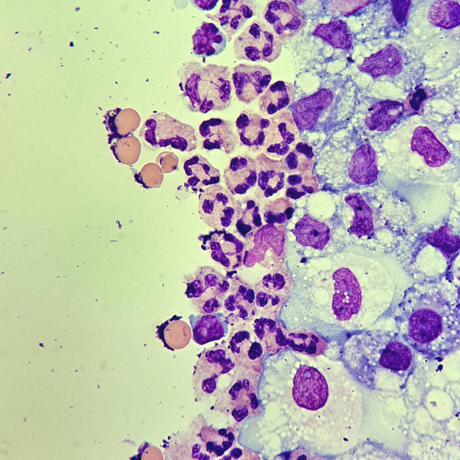
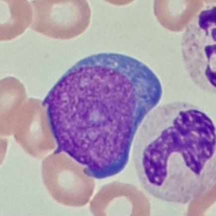
Neutrophils
Cytocentrifugation may make granules in neutrophils appear larger which may be confused for eosinophils. However, they will not have the refractile quality seen in eosinophils or the vibrant reddish-orange coloring.
Lymphocytes
Cytocentrifugation may make lymphocytes appear larger which can be mistaken for blasts if not careful.
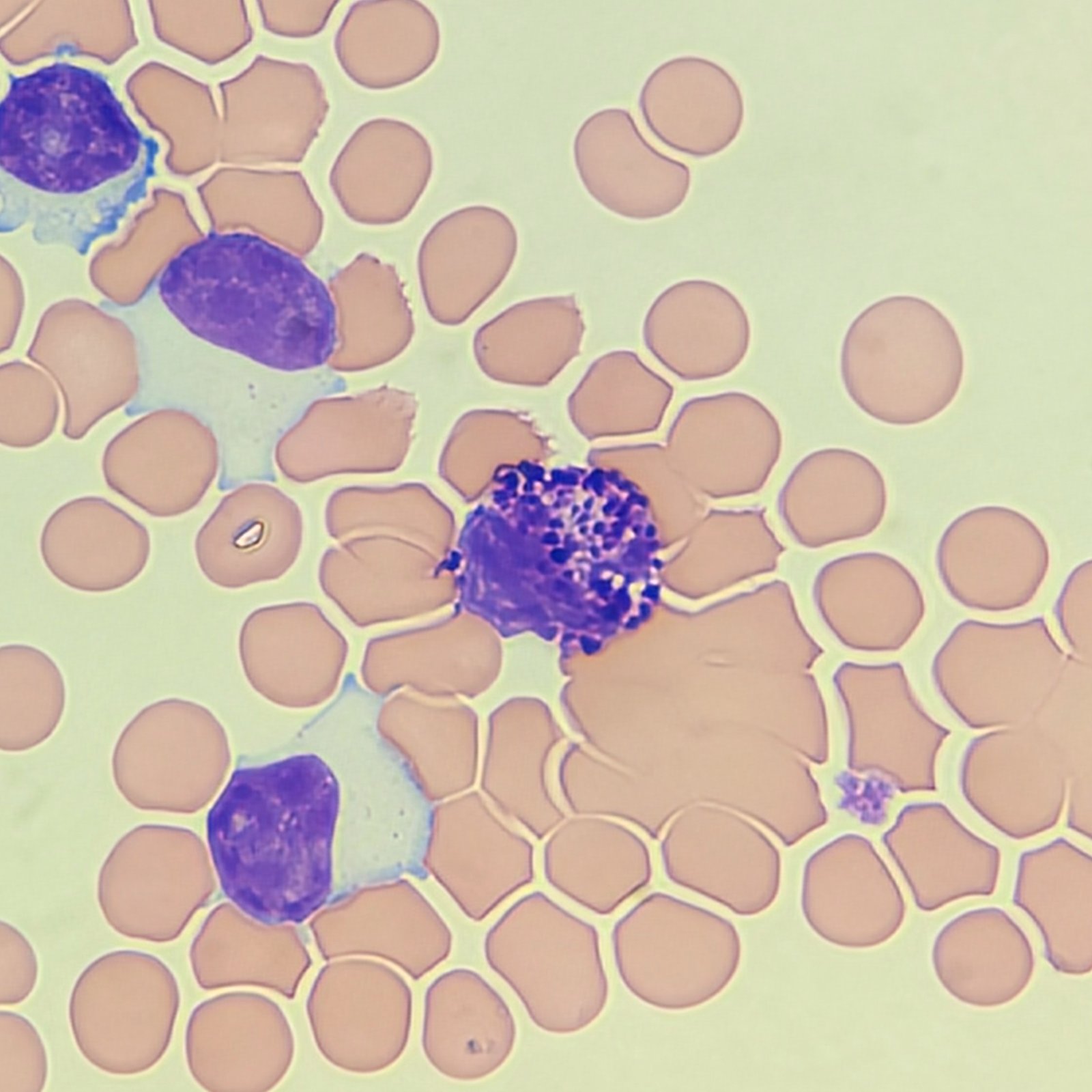
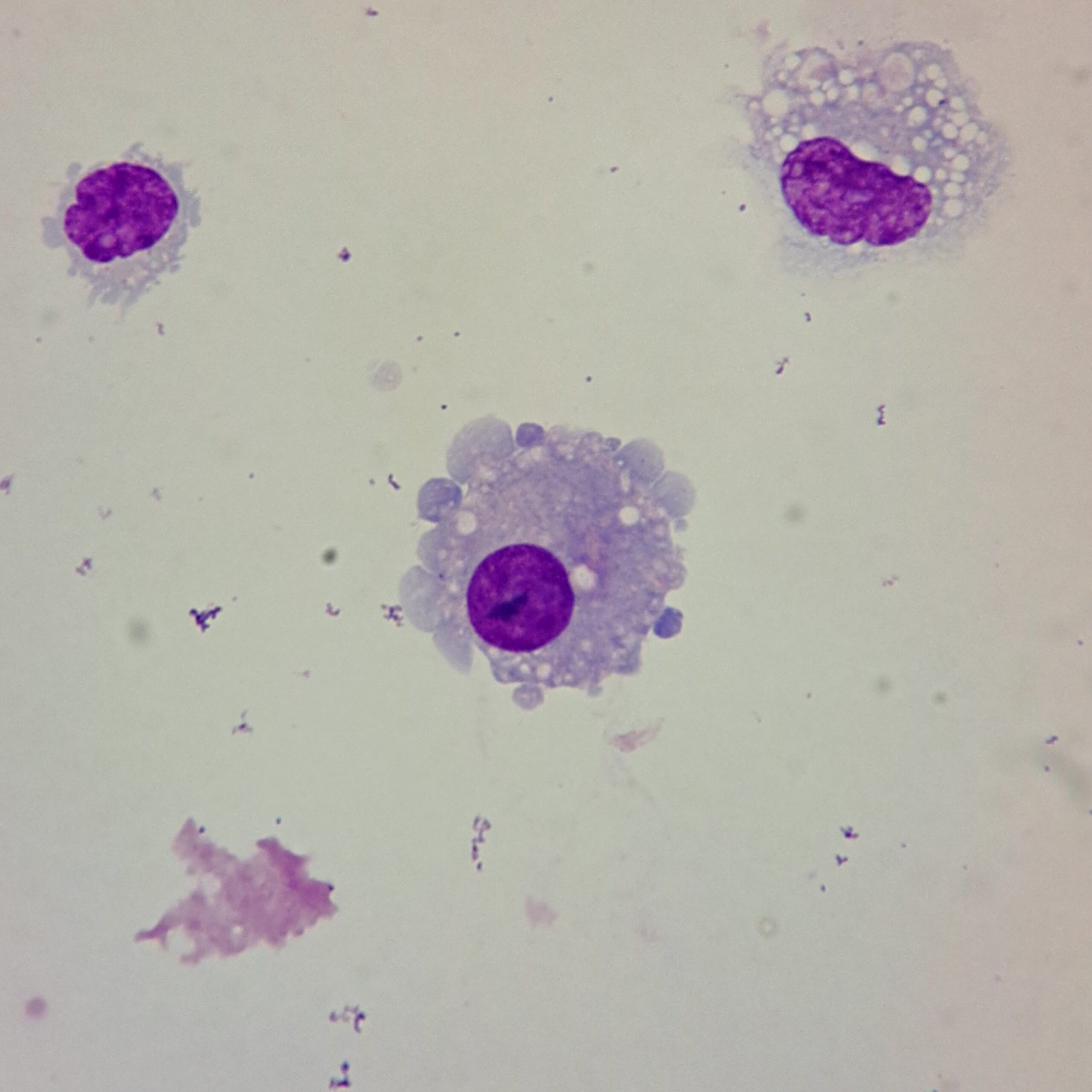
Monocytes / Macrophages
Monocytes in body fluid appear as they do in peripheral blood. Macrophages are monocytes that have differentiated. They are larger and typically have vacuoles.
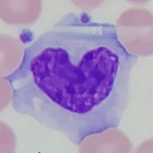
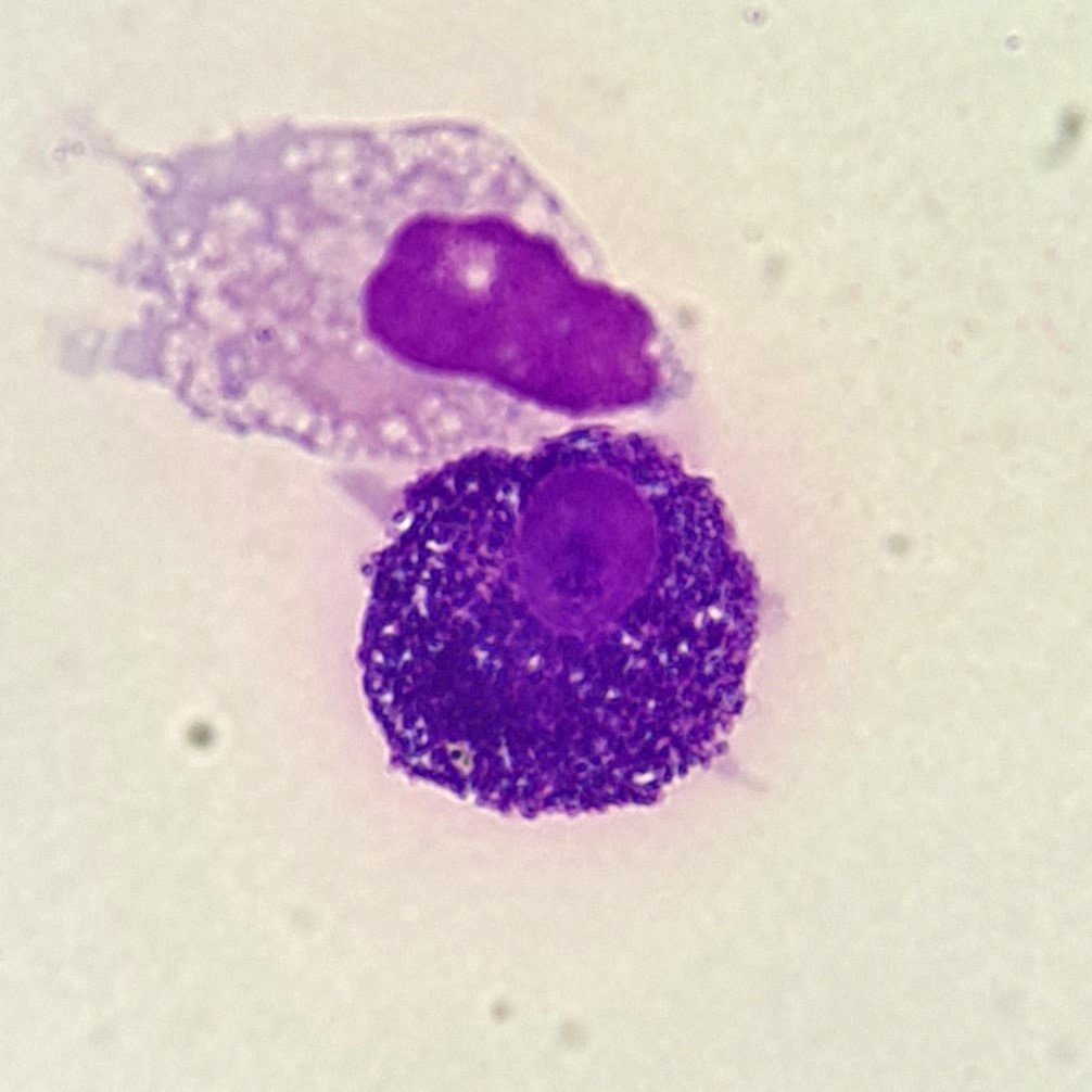
Mast Cells
Larger and more granulated than a basophil but with similar function. Nucleus is round. Cytoplasm is filled with dark blue granules.
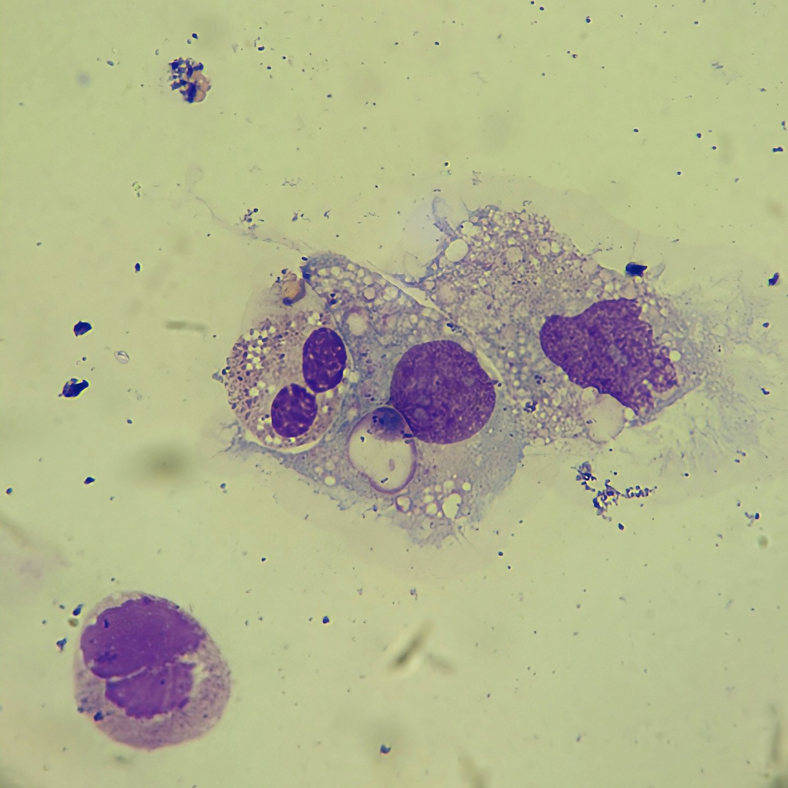
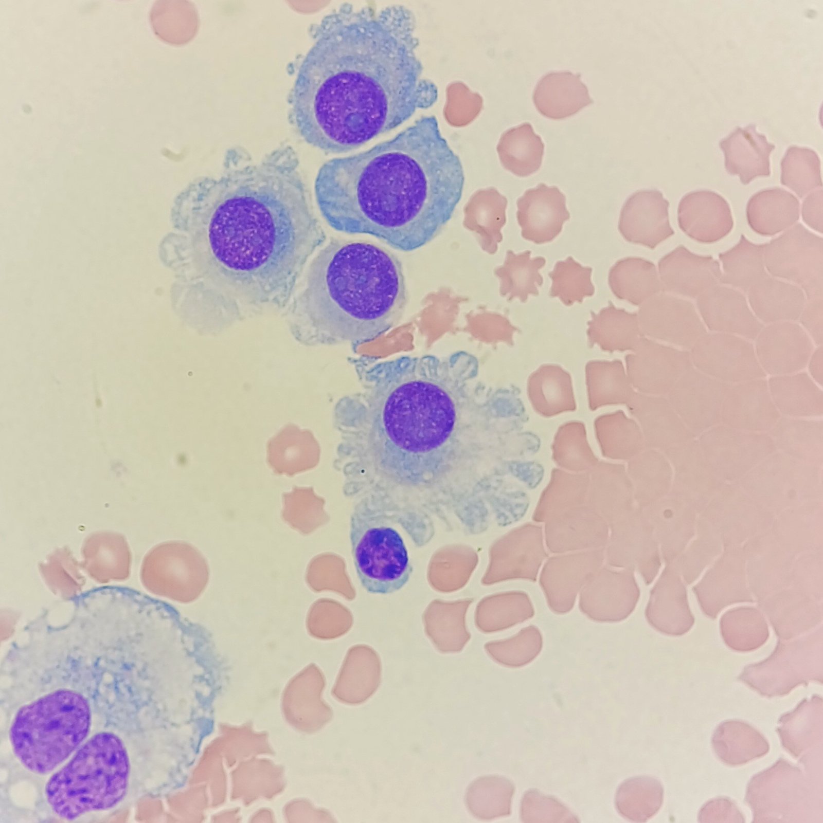
Mesothelial Cells
Lining cells seen in pleural, peritoneal, and pericardial fluids.
Described as having a “fried egg” appearance. Cells may be seen in clumps, but “windows” between cells still allows for individual counting.
Cell may be multinucleated. Nucleus is round to oval with smooth borders and evenly distributed chromatin. Nucleloli are usually present.
Synovial Lining Cells (Synoviocyte)
Lining cell seen in synovial fluid. Visually similar to mesothelial cell.
Nucleus is round to oval with smooth borders. Nucleoli may be present.
Cytoplasm may have irregular edges or vacuoles.
Blast Cells
Cytocentrifugation may exaggerate cellular features, such as larger nucleoli or irregular nuclear shape.
Chromatin is fine with one or more nucleoli.
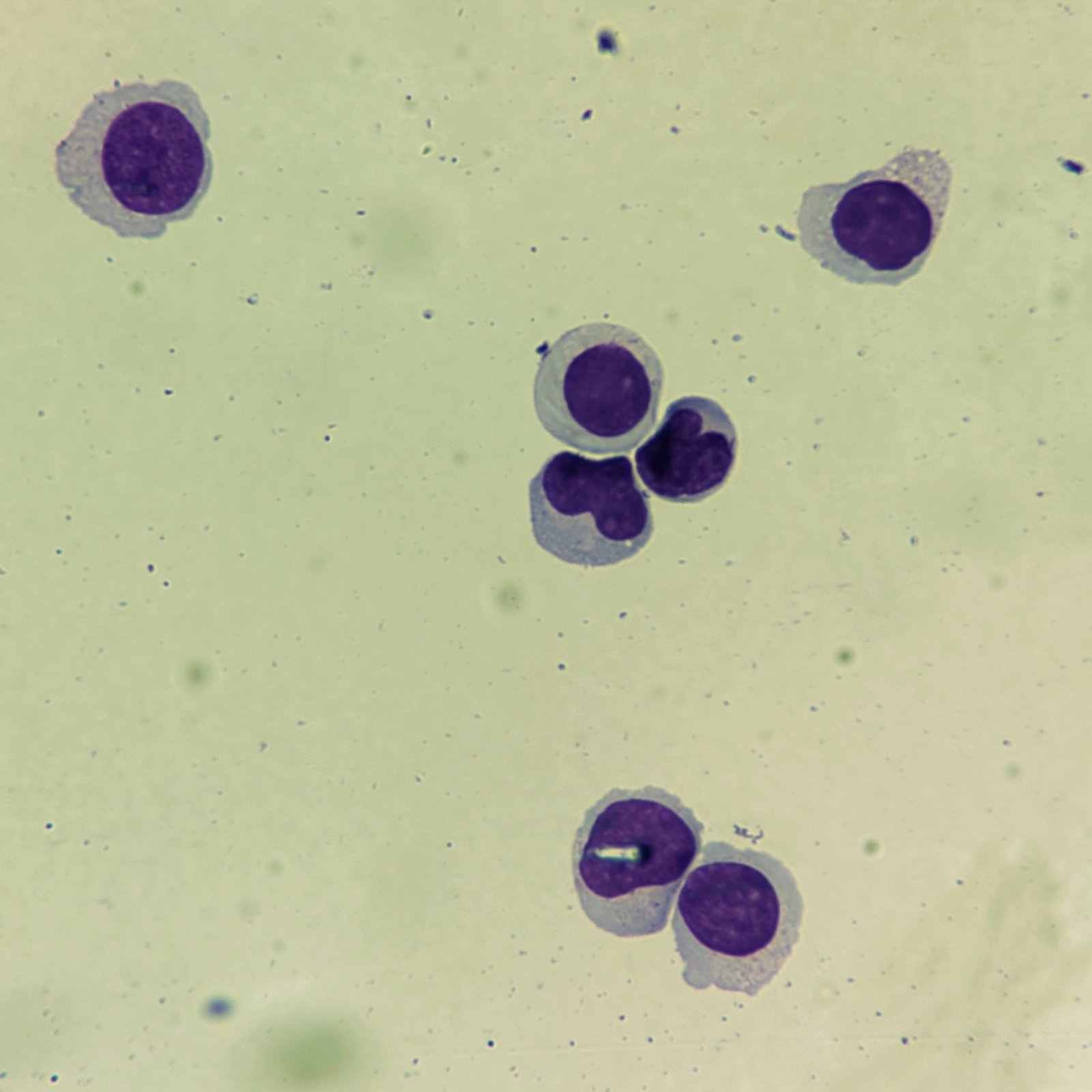
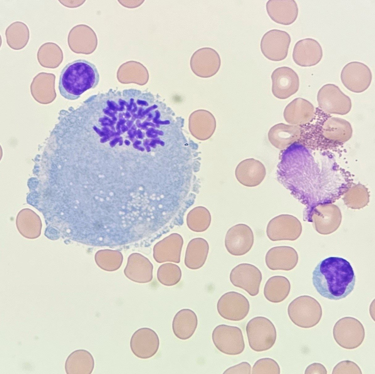
Mitotic Figures
Chromosomal structures are visible in the nucleus, which can cause a daisy-like appearance.
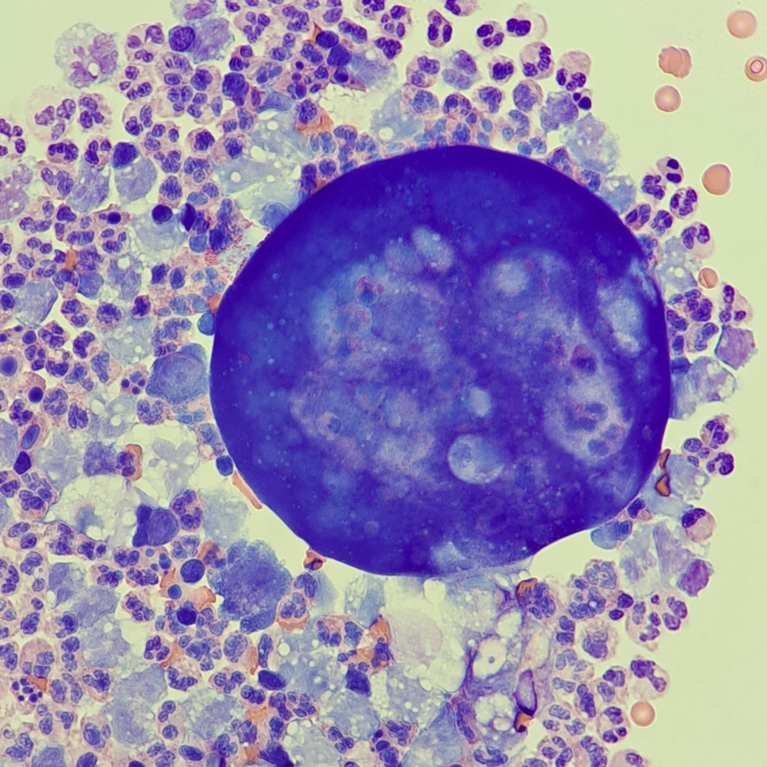
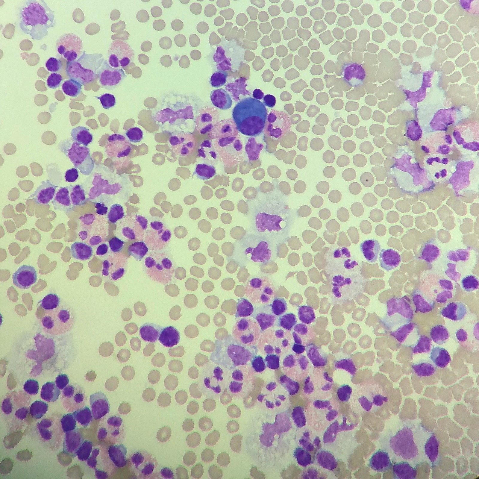
Malignant Cells
Appearance of malignant cells can vary widely. However, they commonly have one or more of the following features: Cells clumping together with no discernable borders between or one giant cell with many nuclei, Dark-staining, 3-D appearance, irregular nulei or chromatin pattern, etc.