Malignant cells may be found in body fluids due to blood cancers or other cancers that have metastasized. These slides should be sent to the pathologist for examination and further study.
Appearance
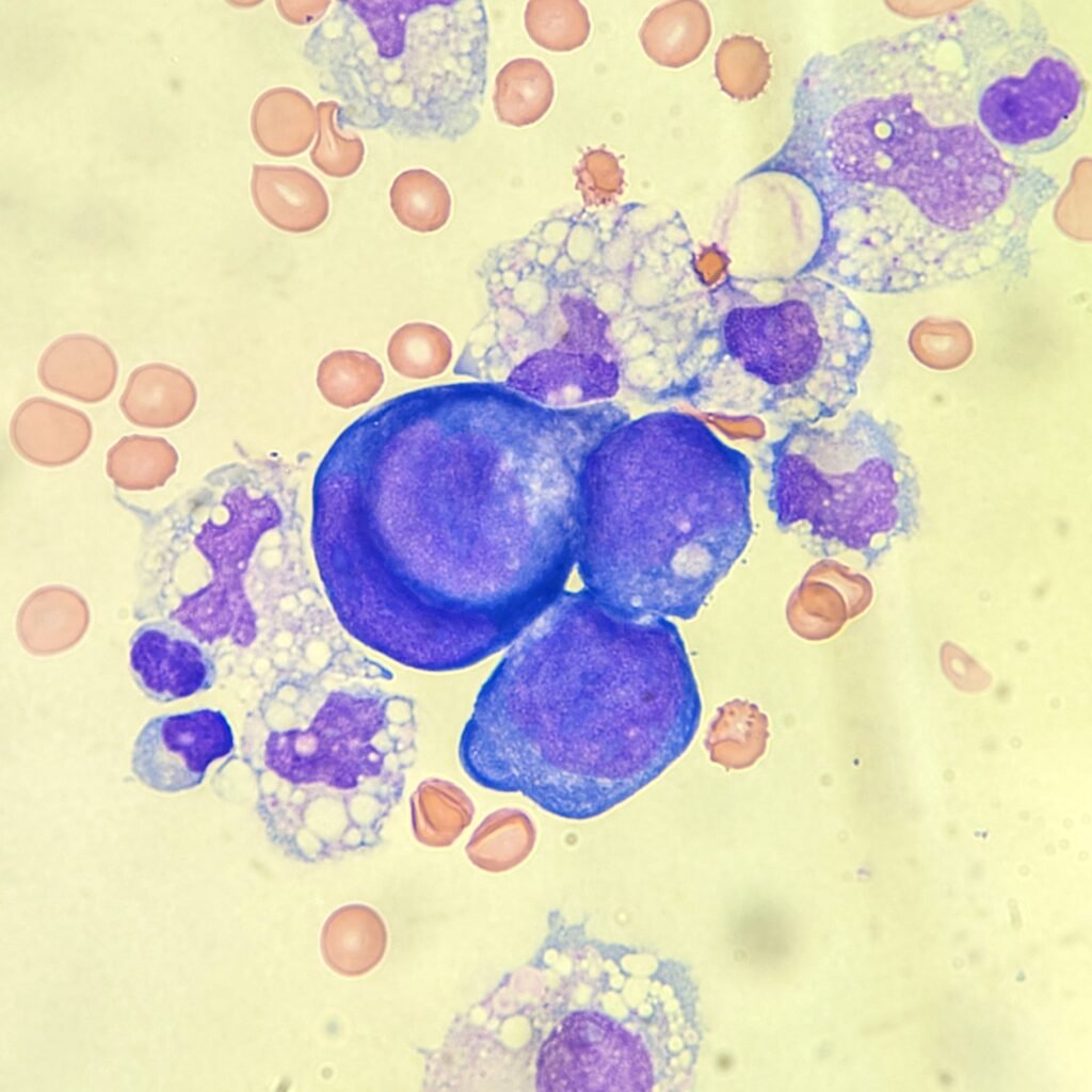
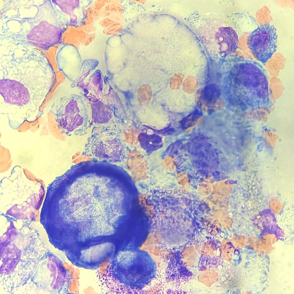
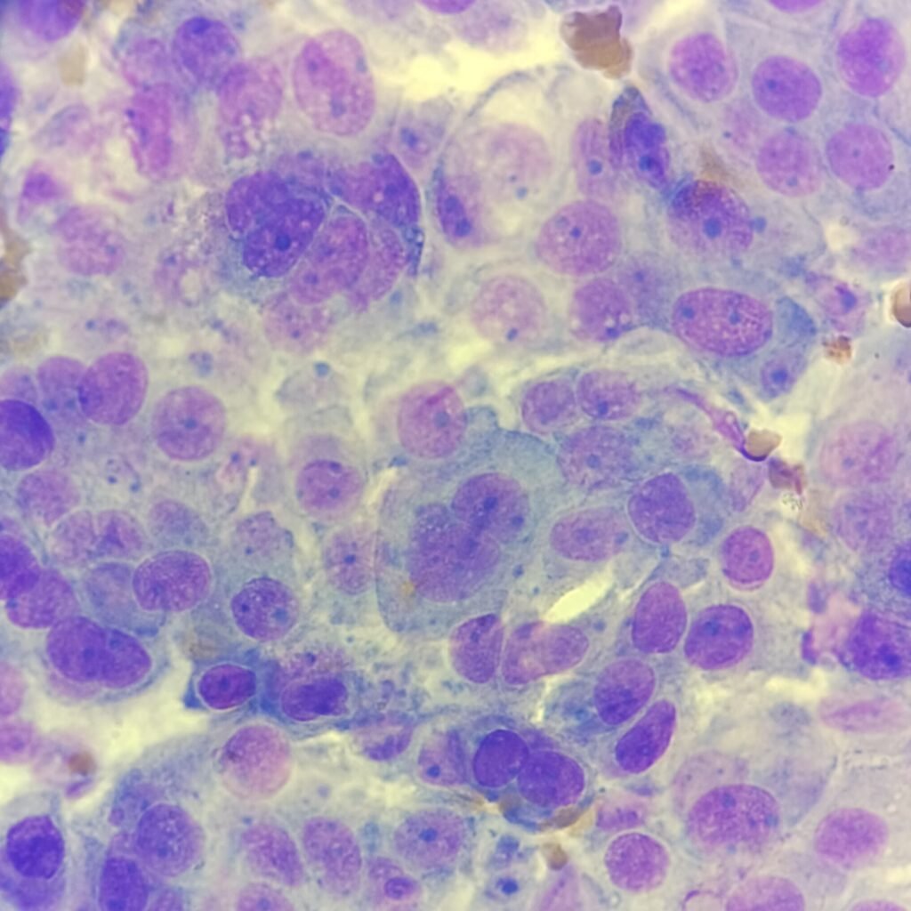
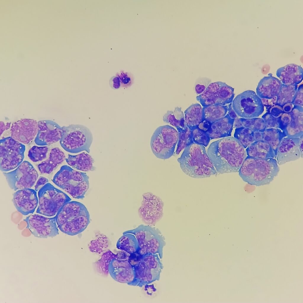
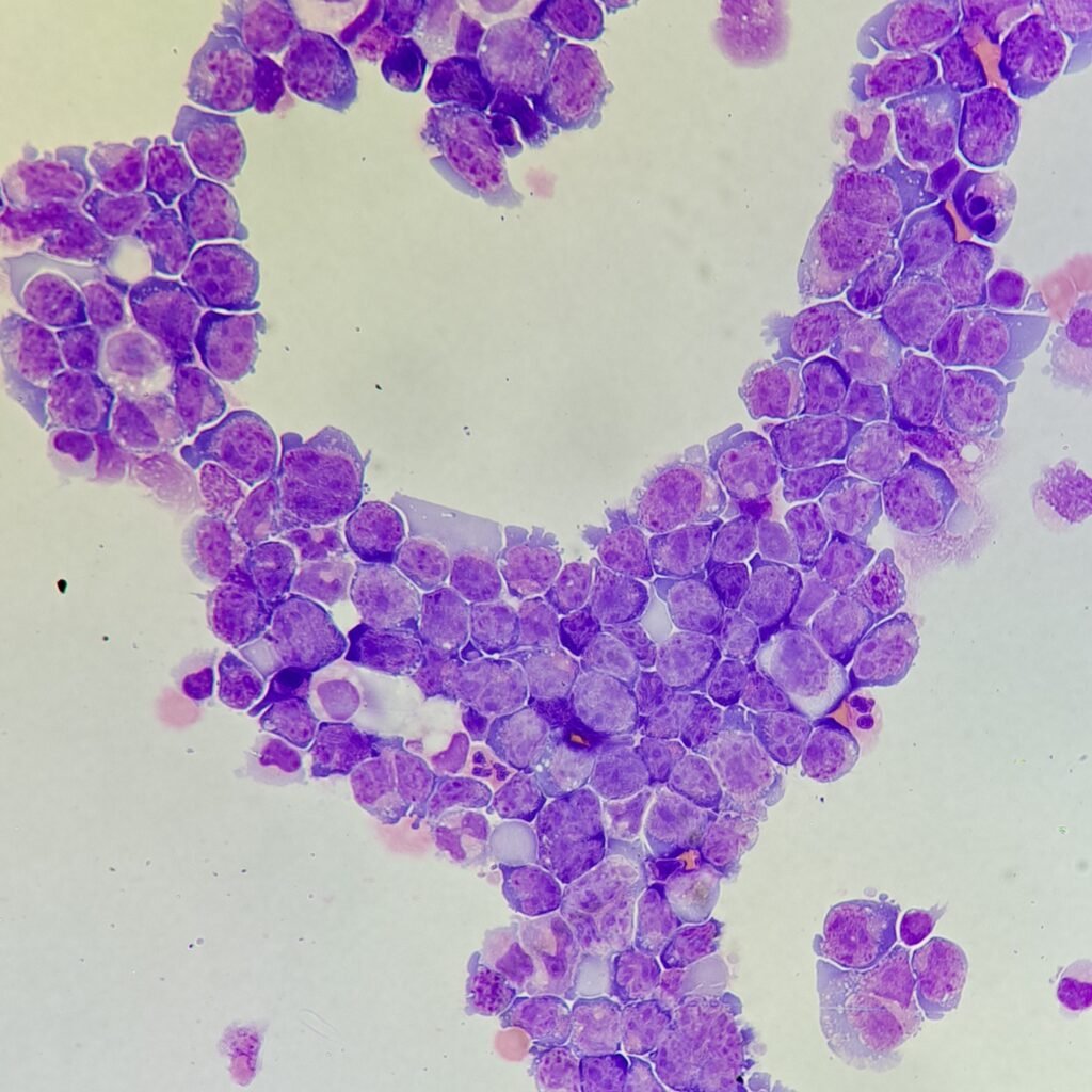
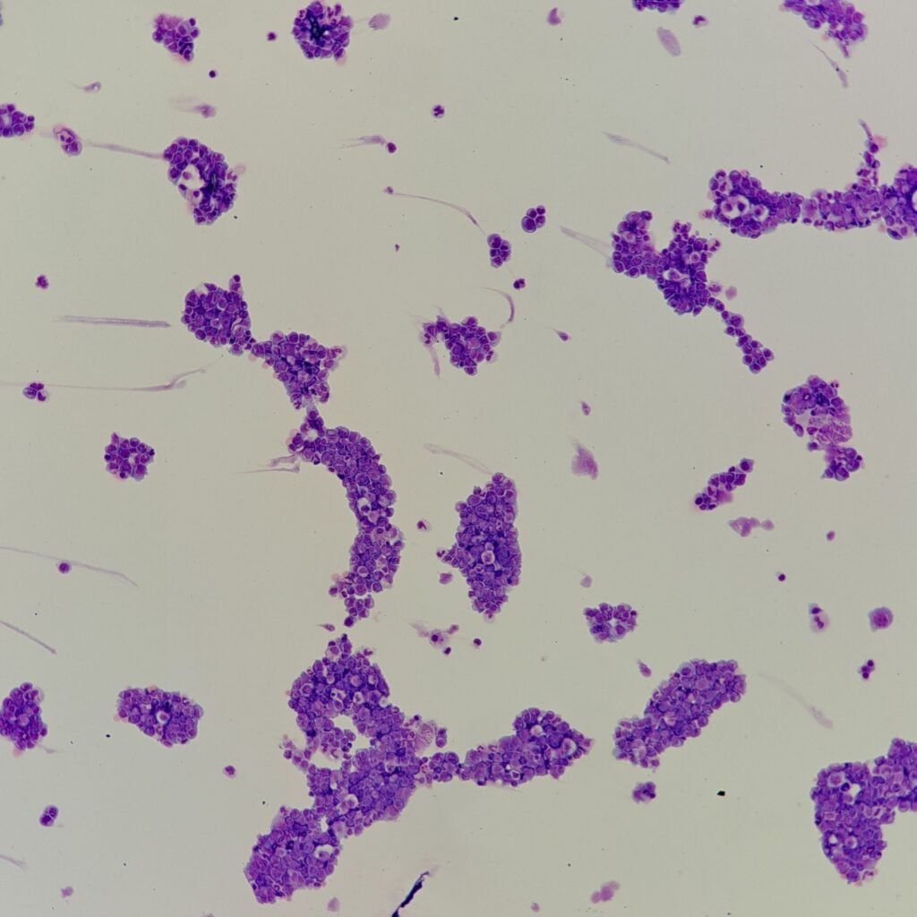
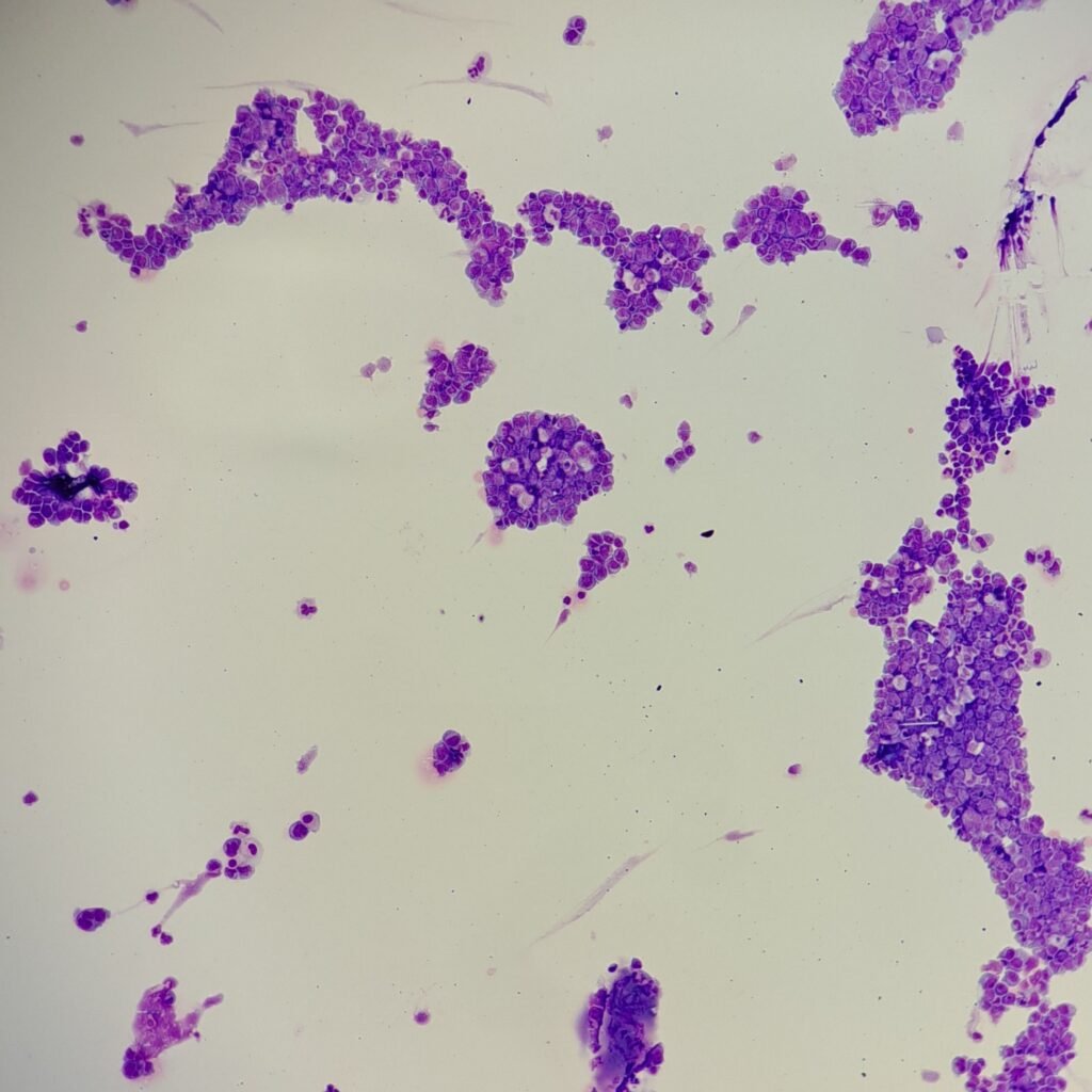
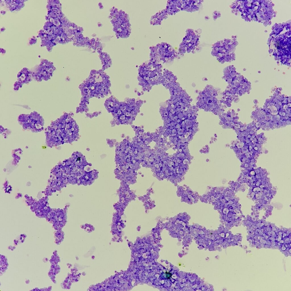
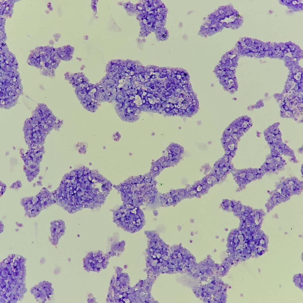
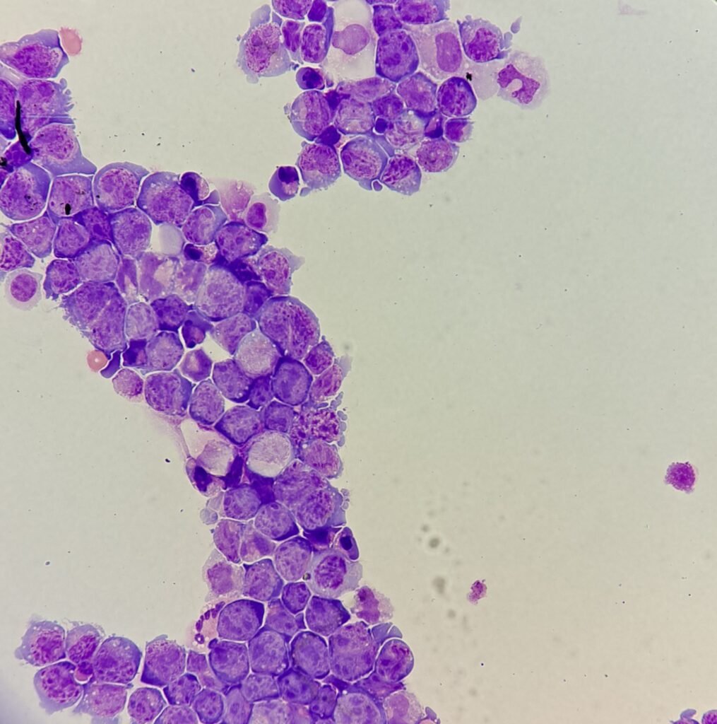
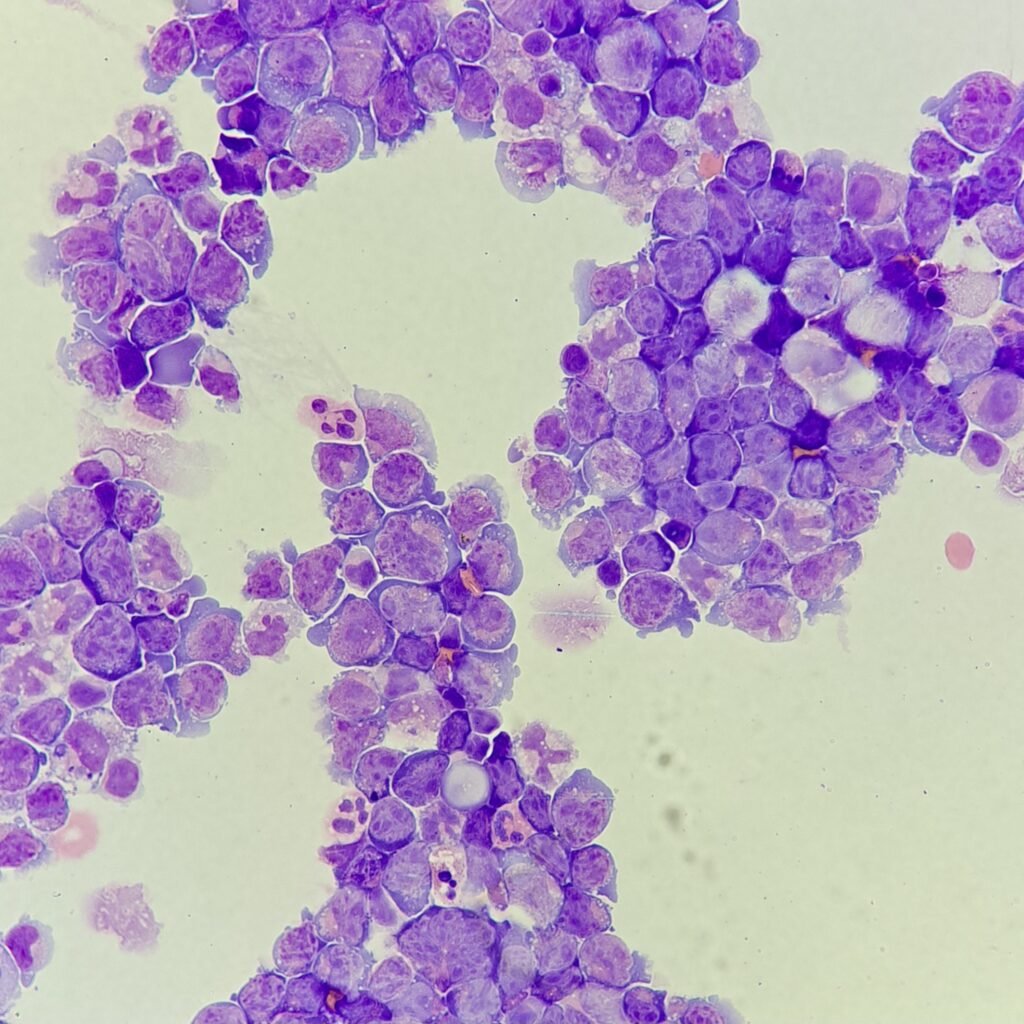
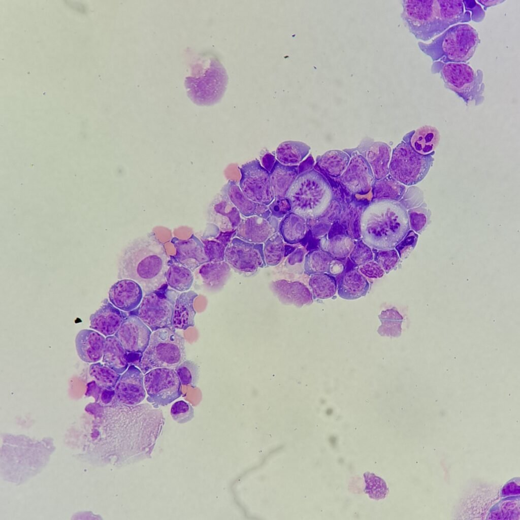
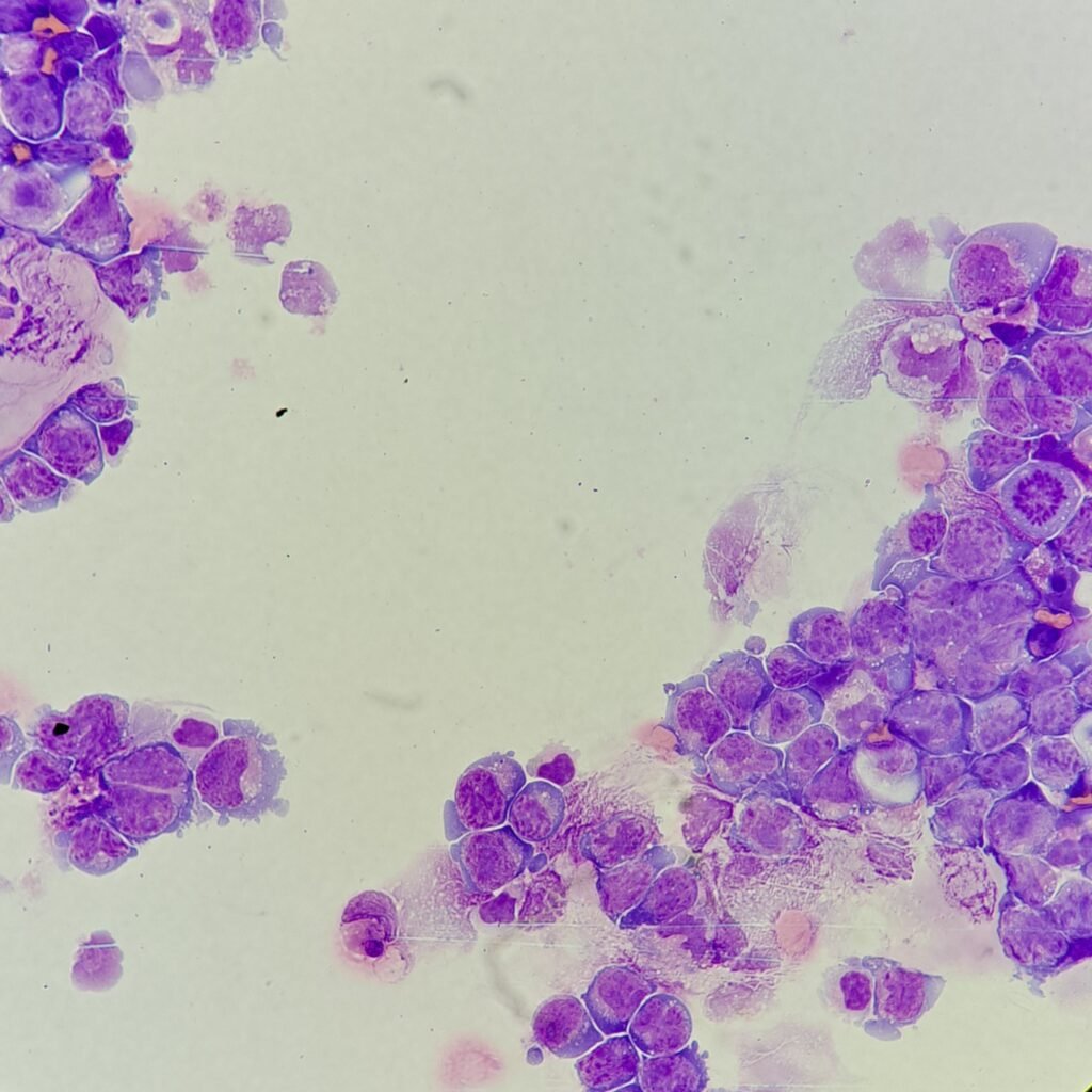
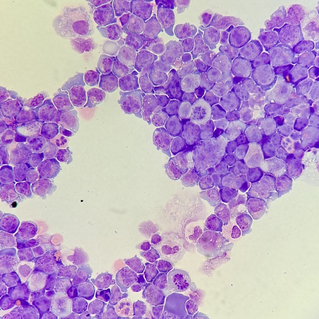
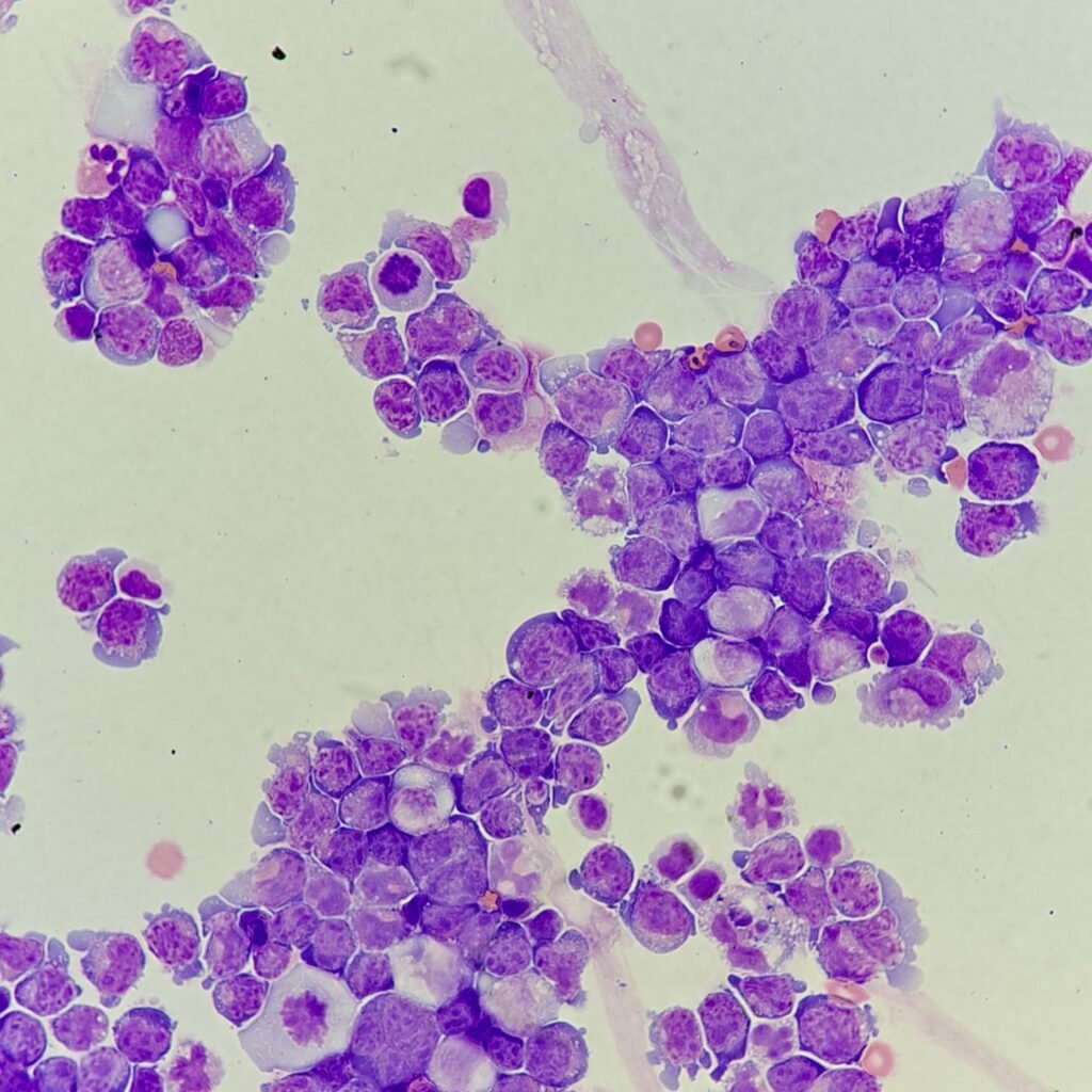
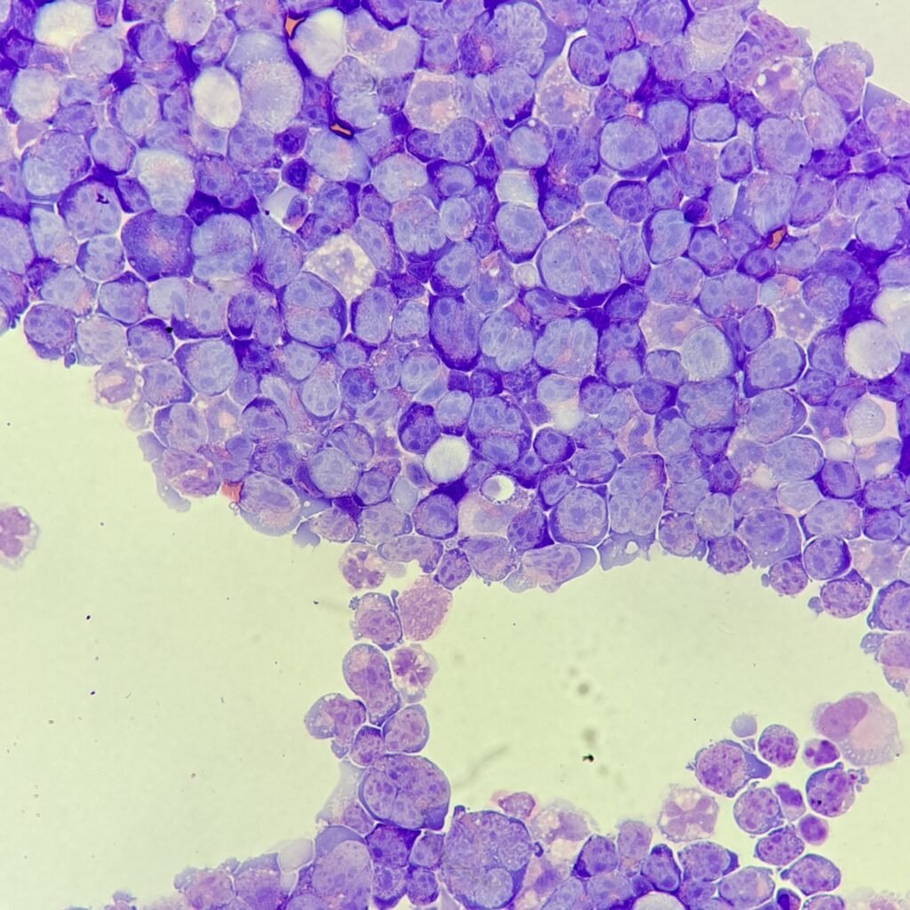
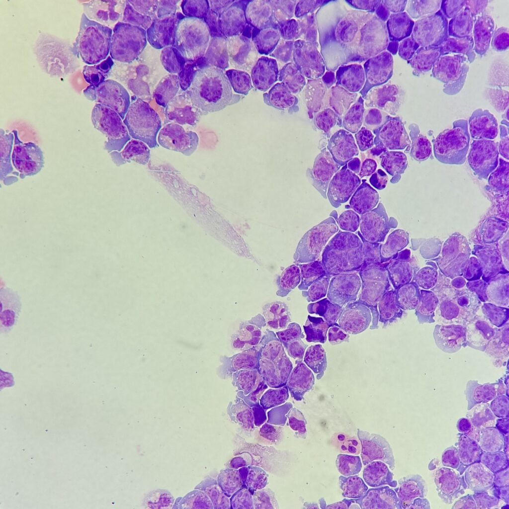
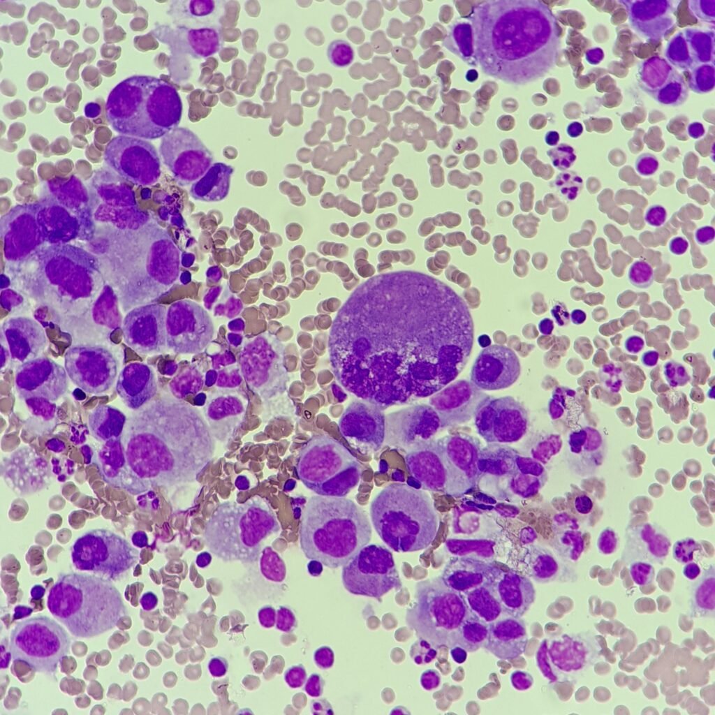
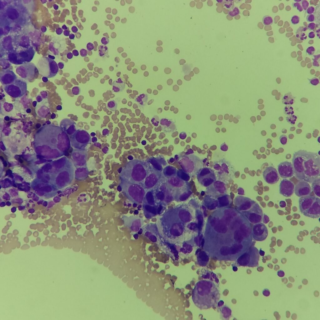
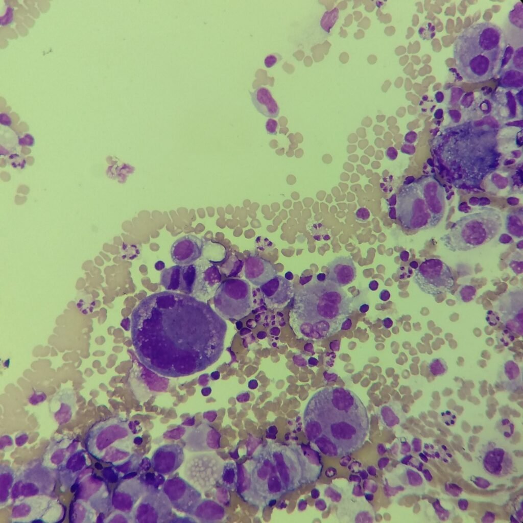
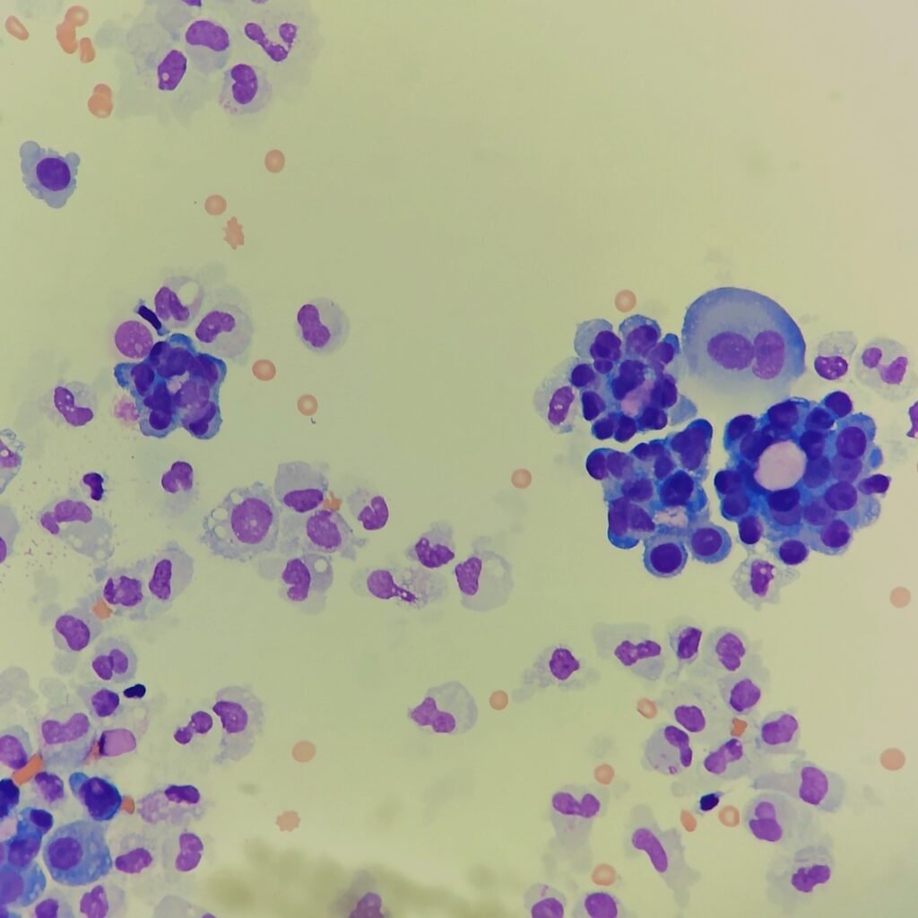
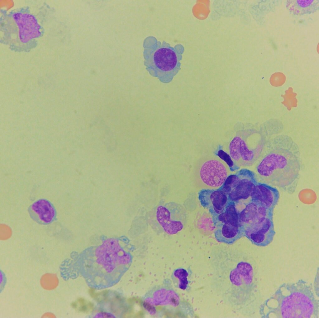
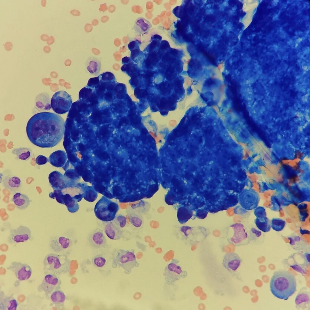
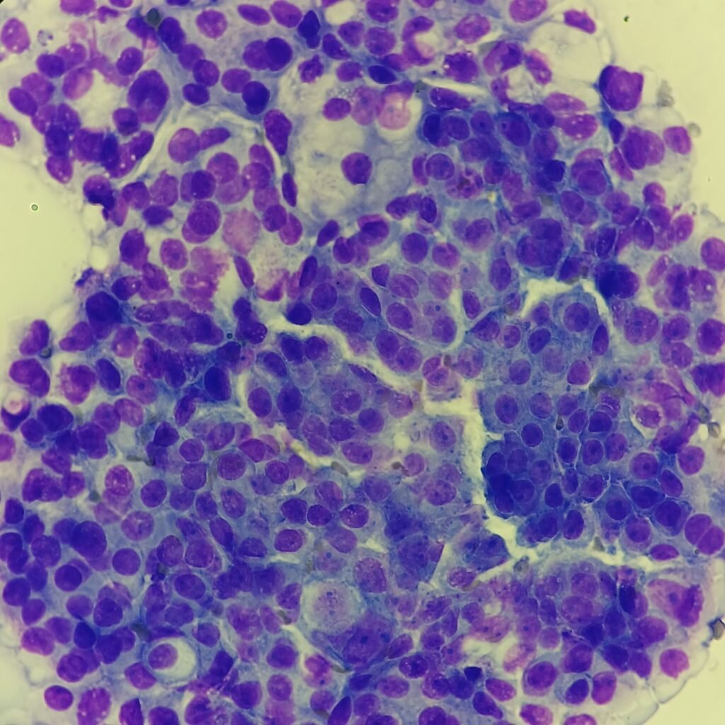
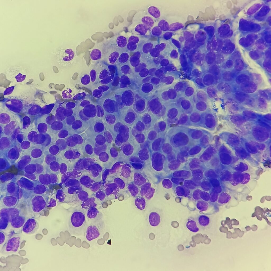
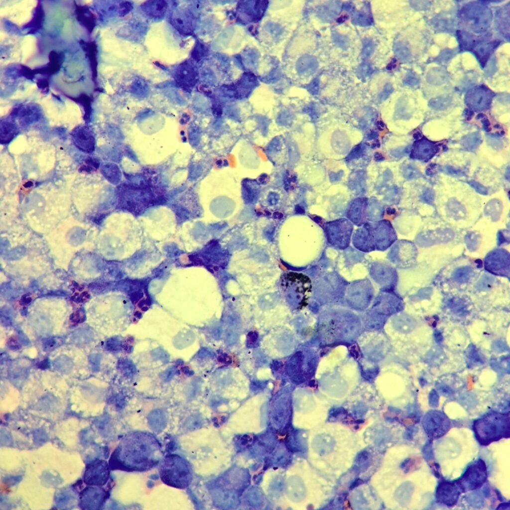
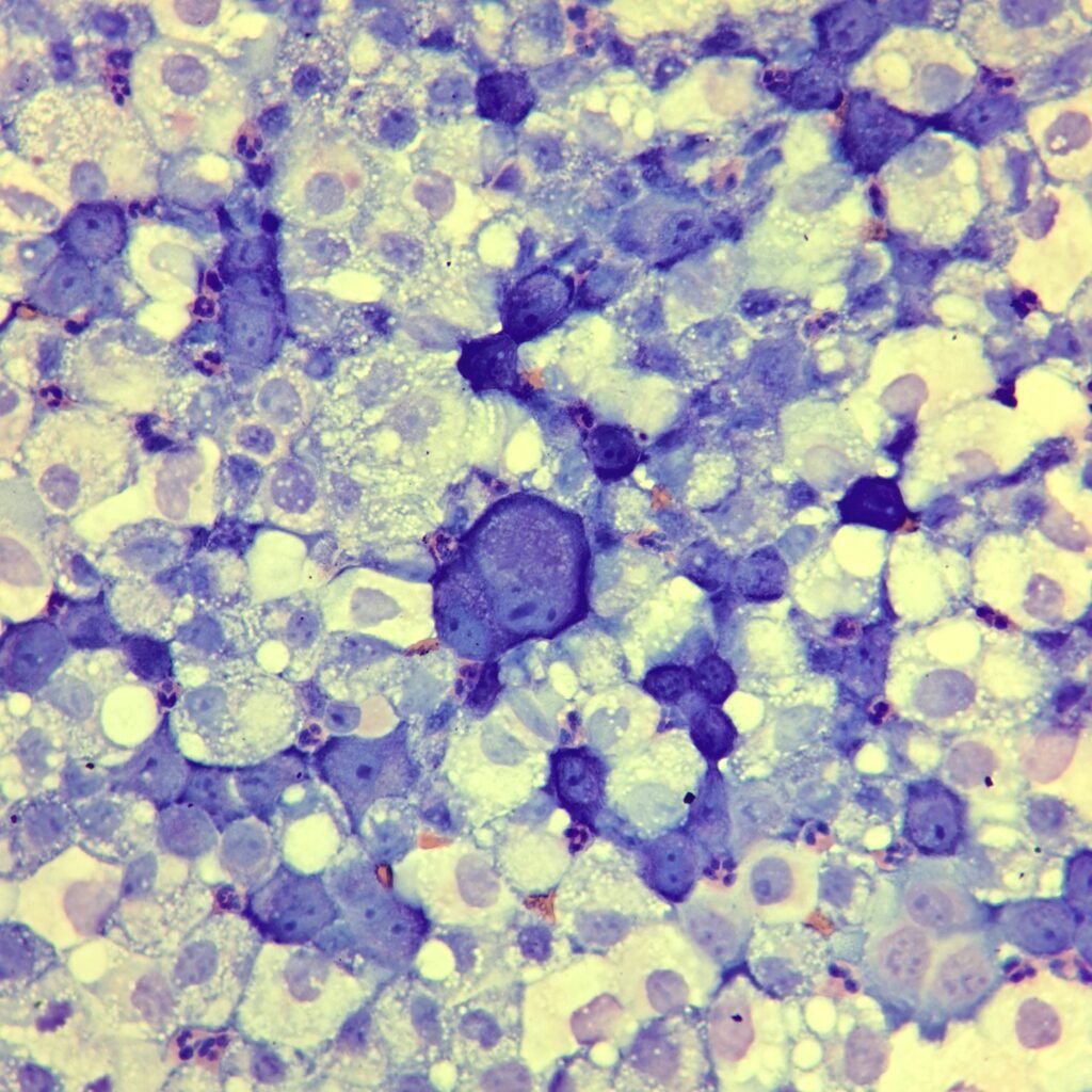
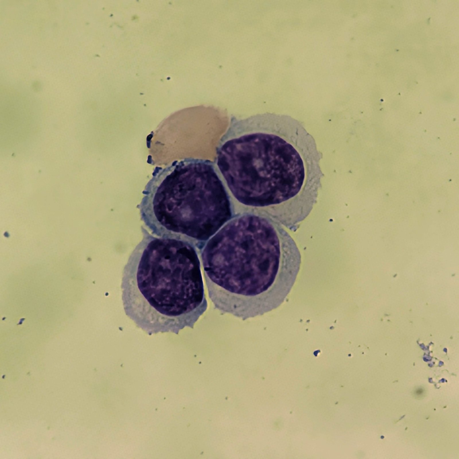
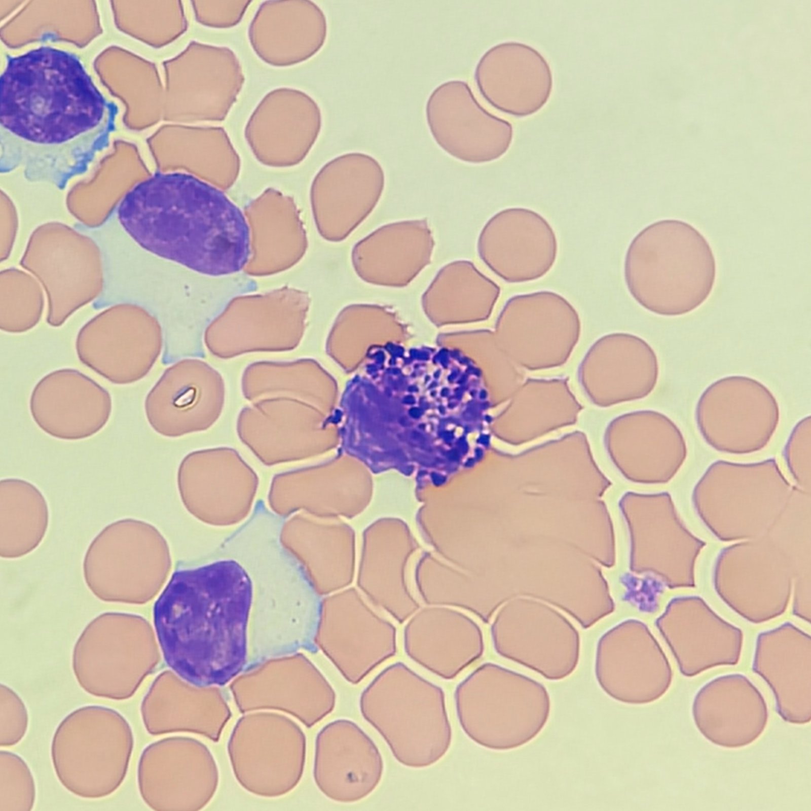
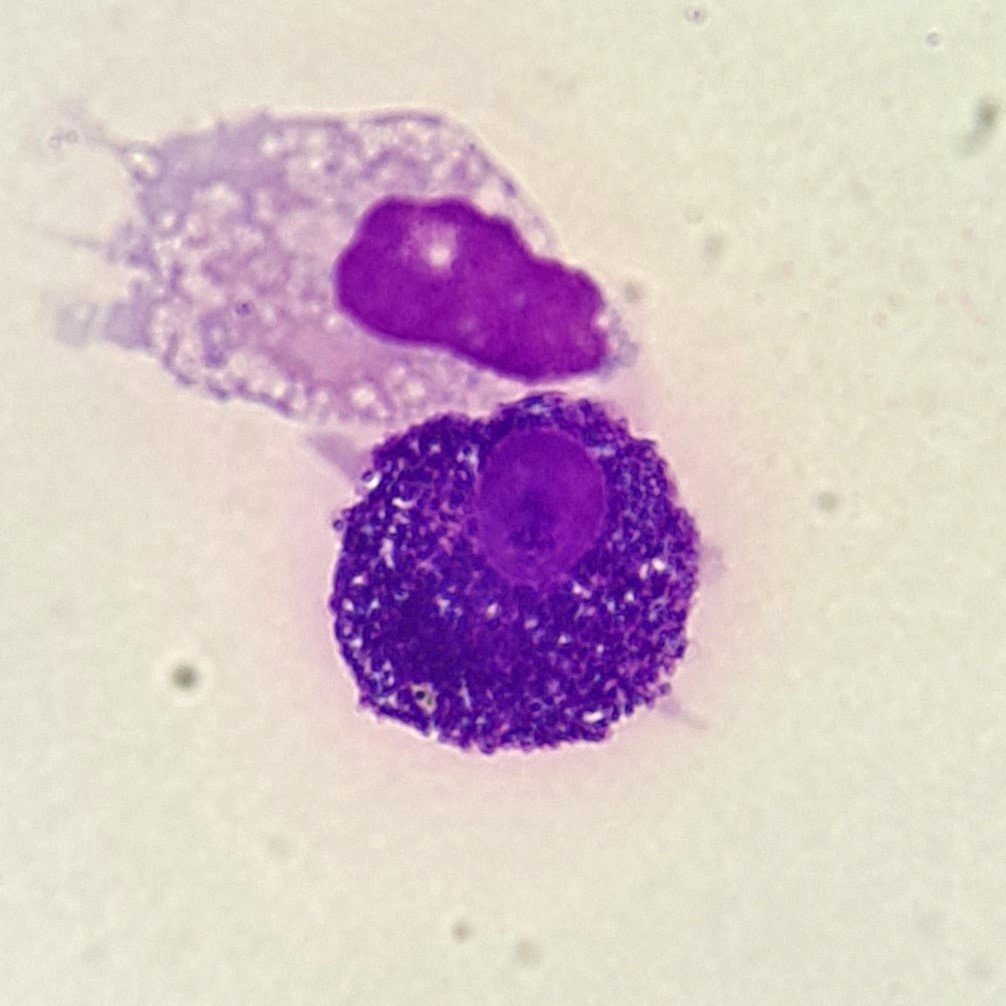
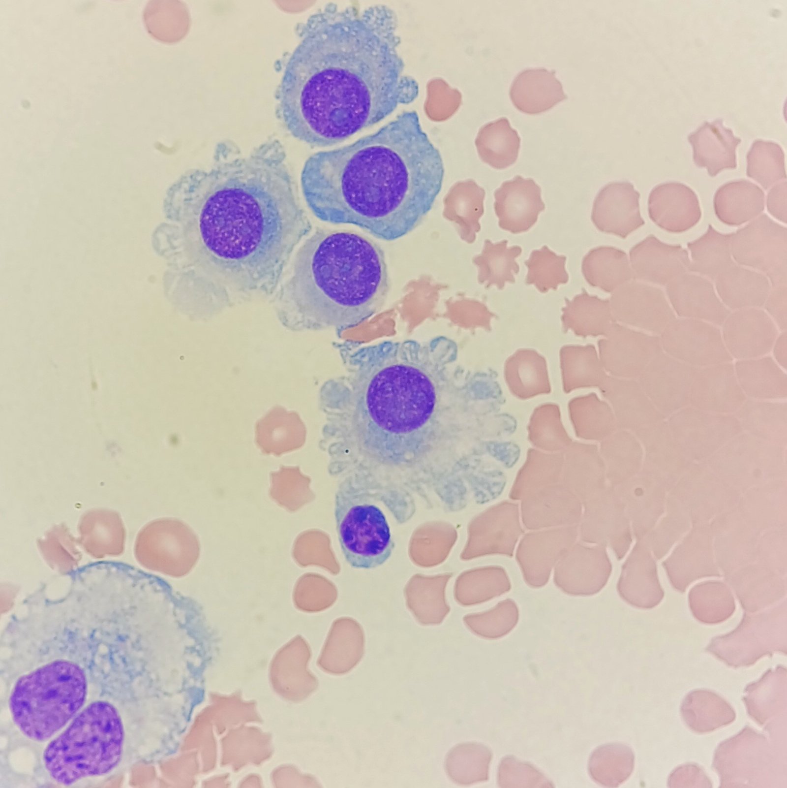
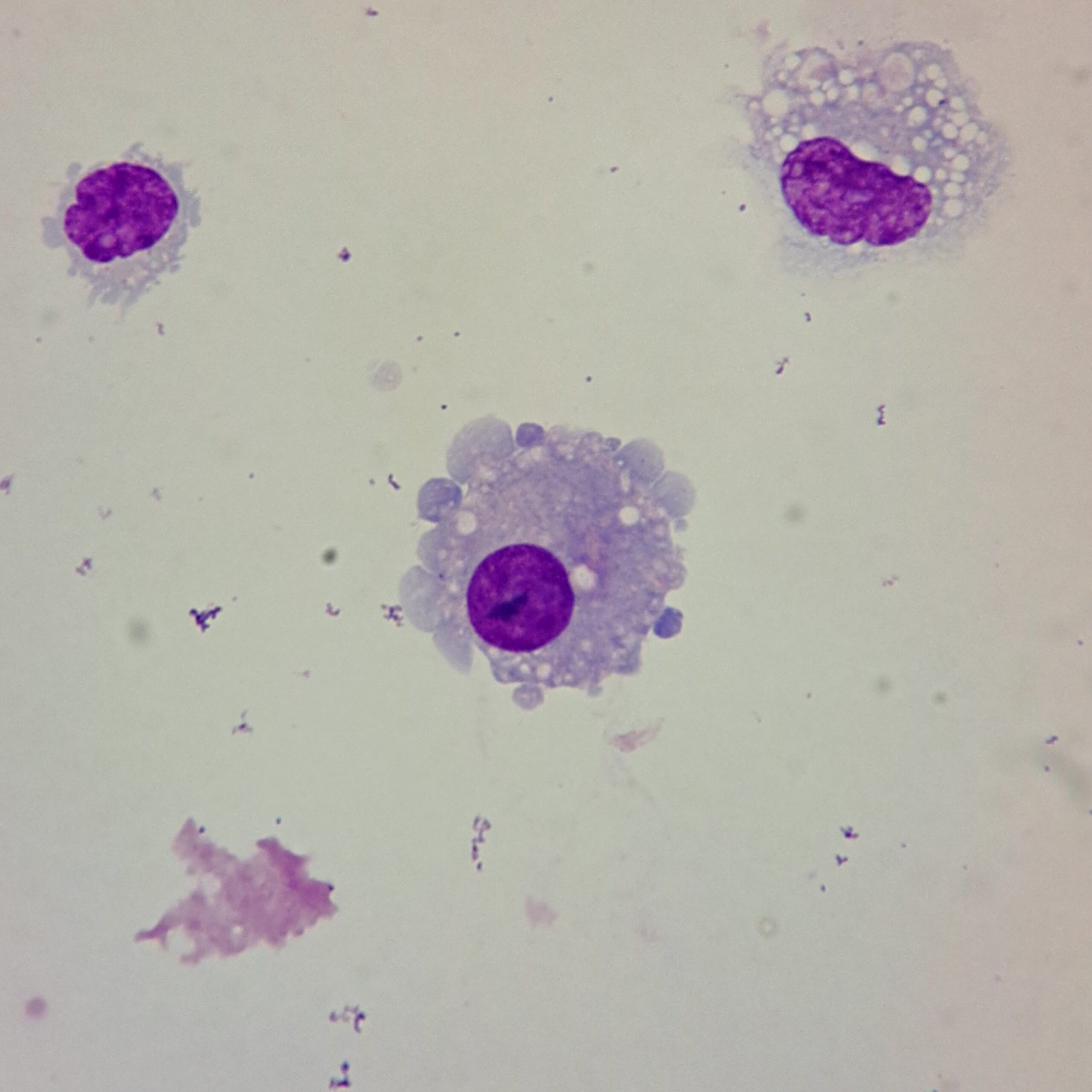
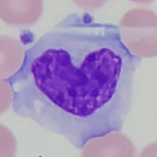
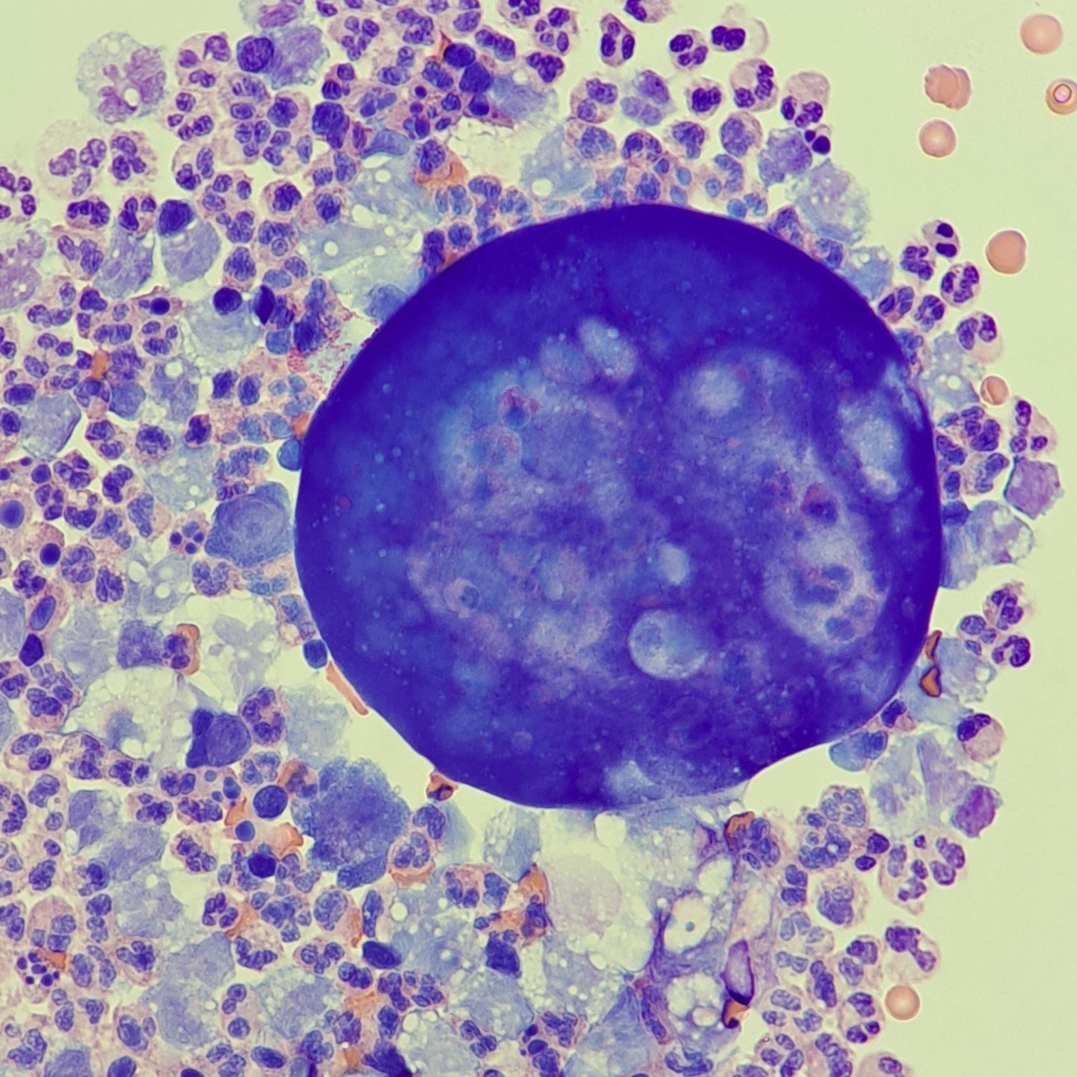
Appearance of malignant cells can vary widely. However, they commonly have one or more of the following features: Cells clumping together with no discernable borders between or one giant cell with many nuclei, Dark-staining, 3-D appearance, irregular nuclei or chromatin pattern, etc.
Malignant cells are often apparent on low power due to these distinctive features. Because these cells may be few in number or clump together, it is important to scan the cytocentrifuge slide for the presence of them.
Lookalikes
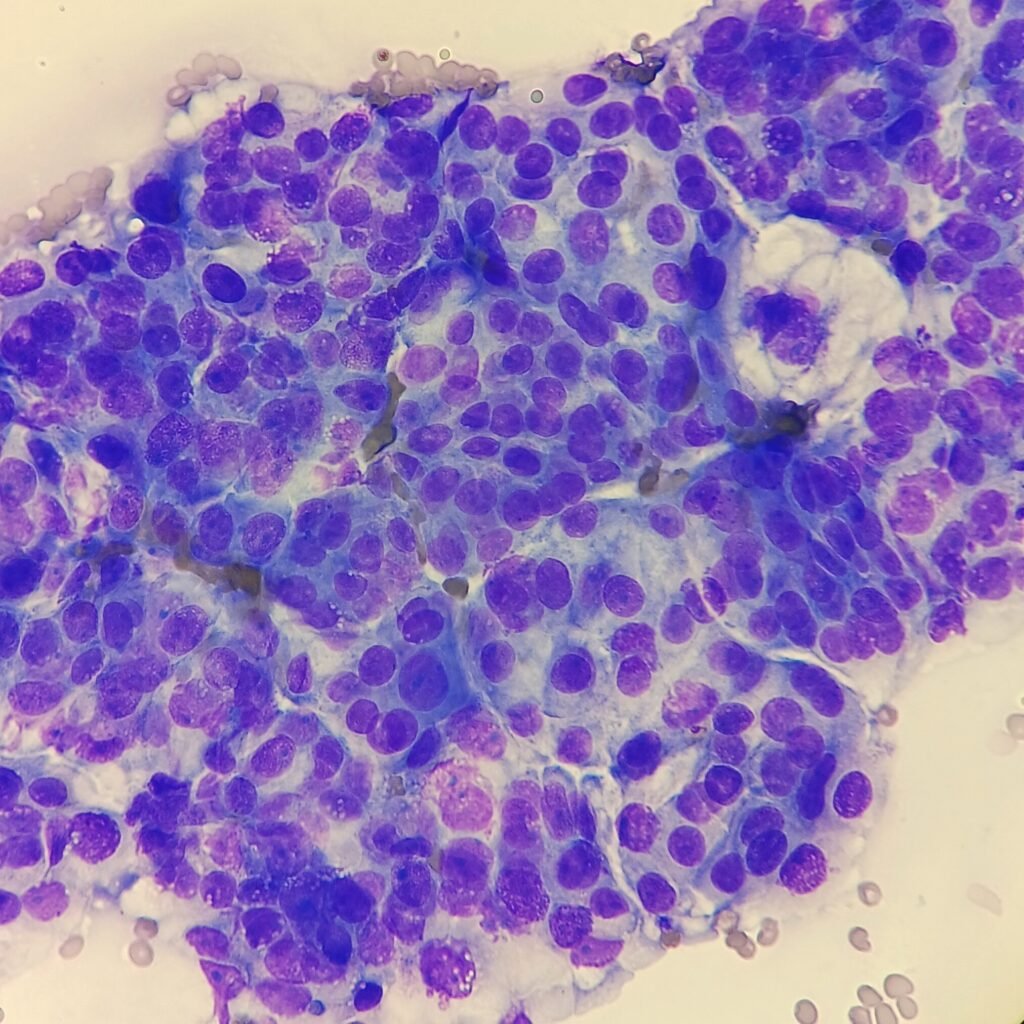
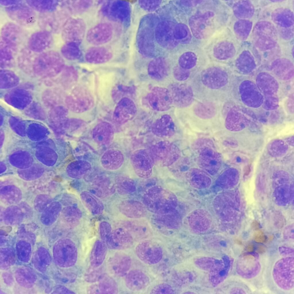
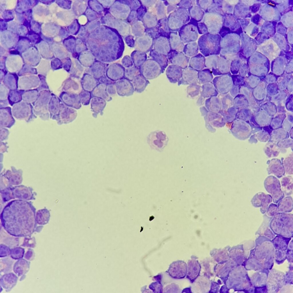
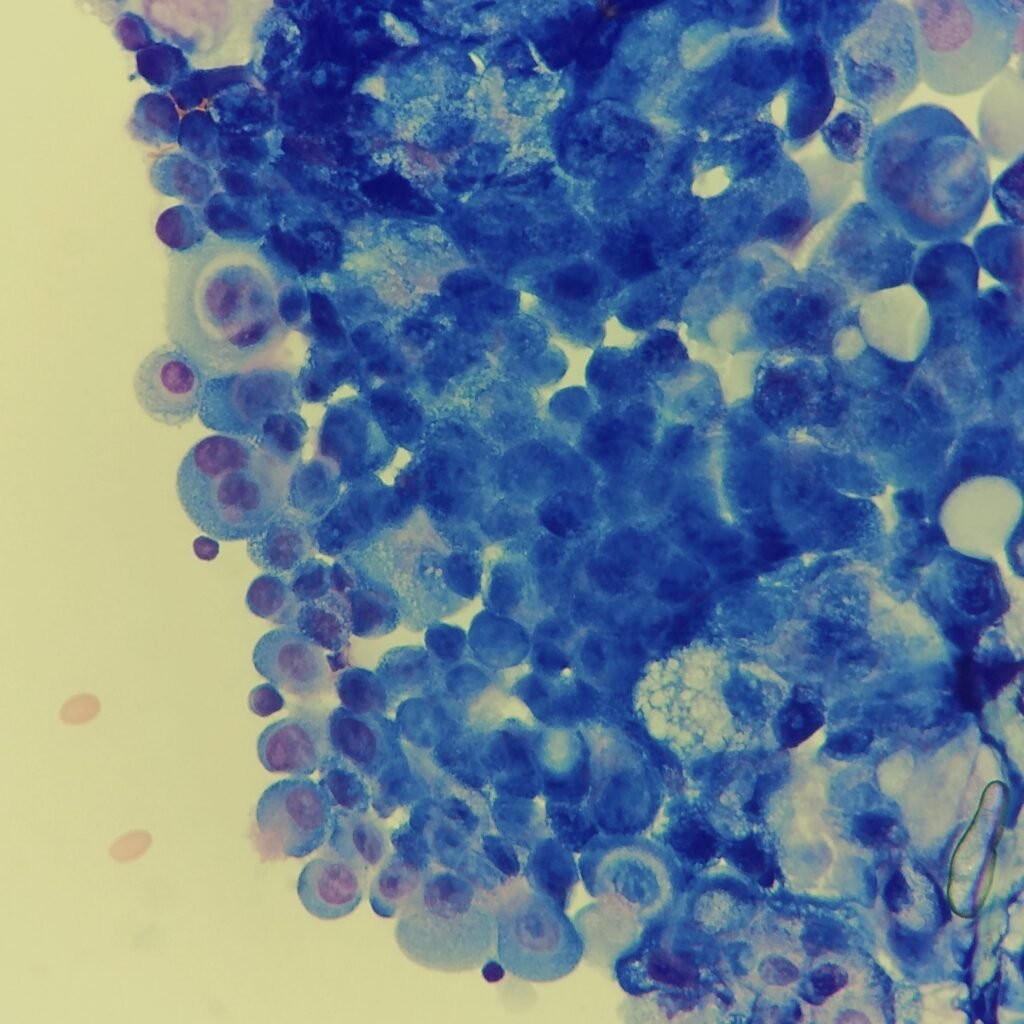
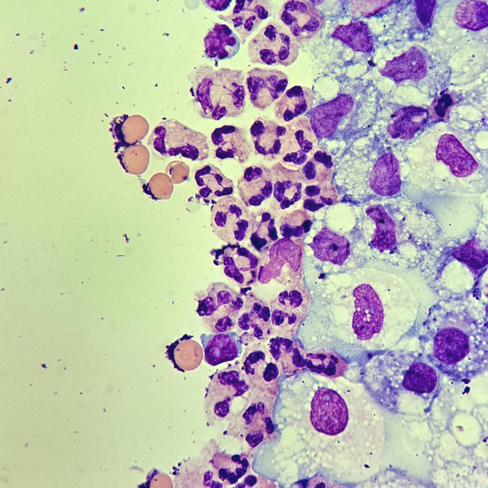
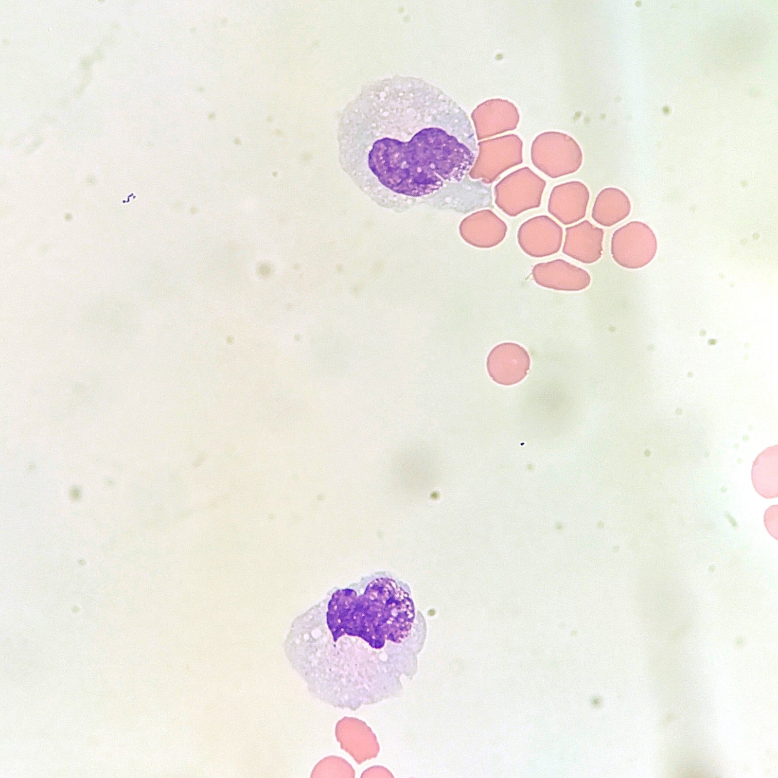
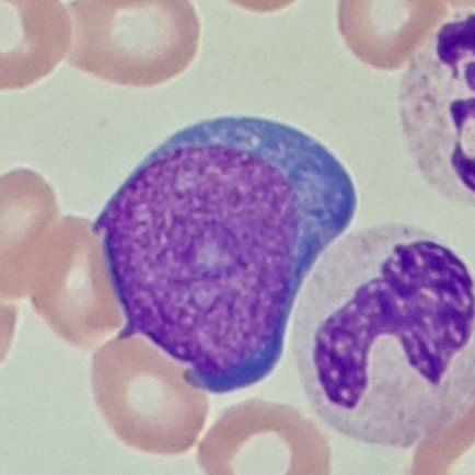
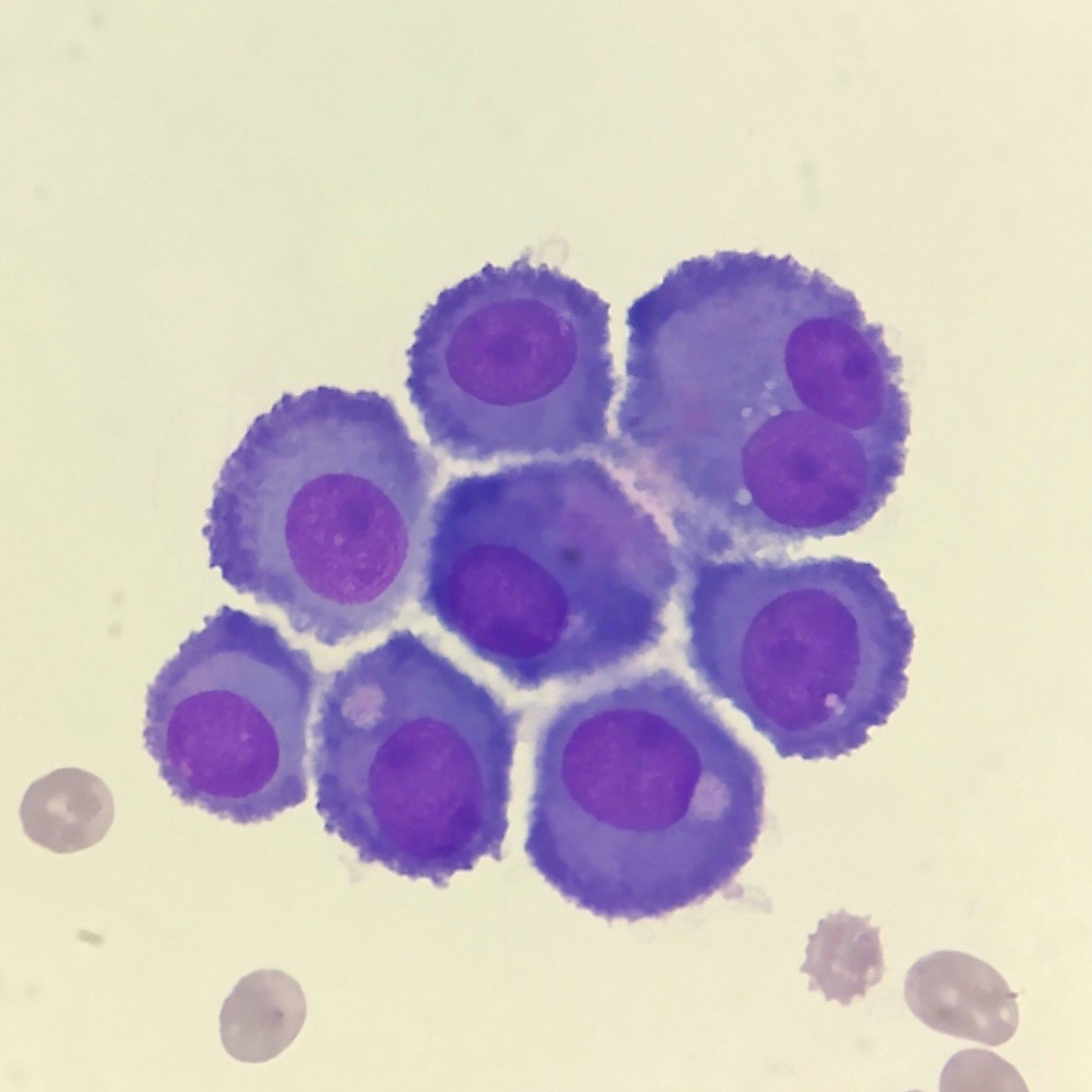
Both malignant cells and mesothelial cells can clump together. However, mesothelial cells will have windows between the cells that allow for proper differentiation. Reactive mesothelial cells can be especially difficult to distinguish from malignancy.
Gallery