Mesothelial cells are lining cells seen in pleural, peritoneal, and pericardial fluids.
Appearance
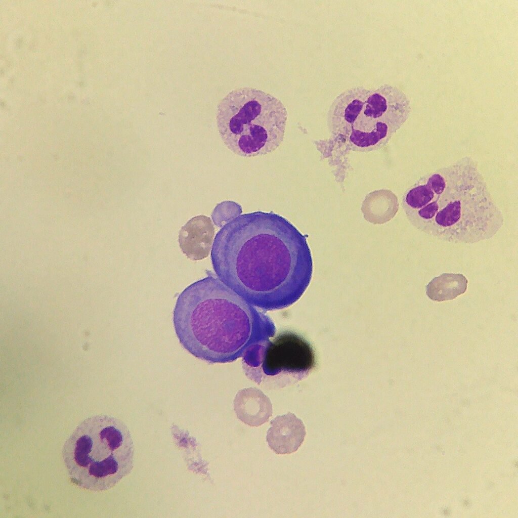
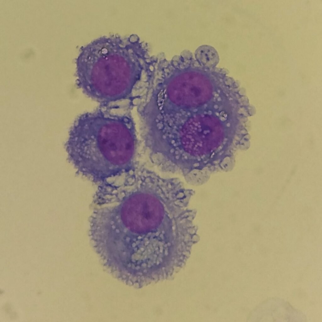
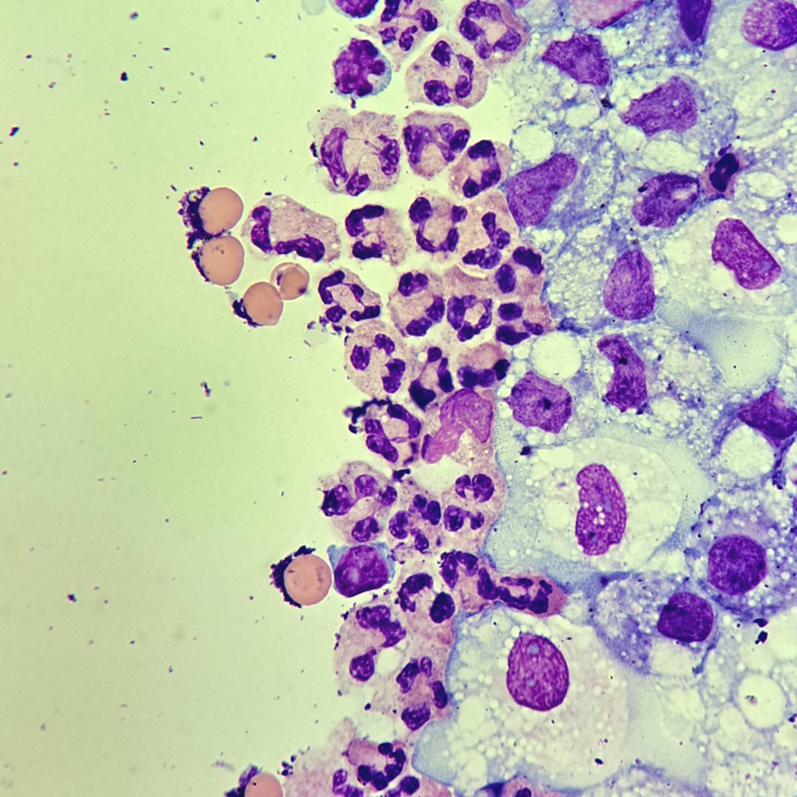
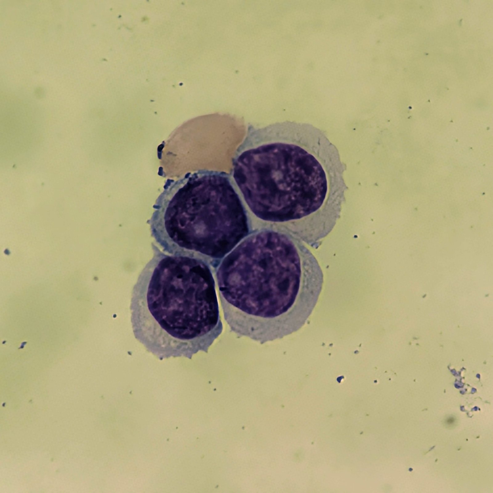
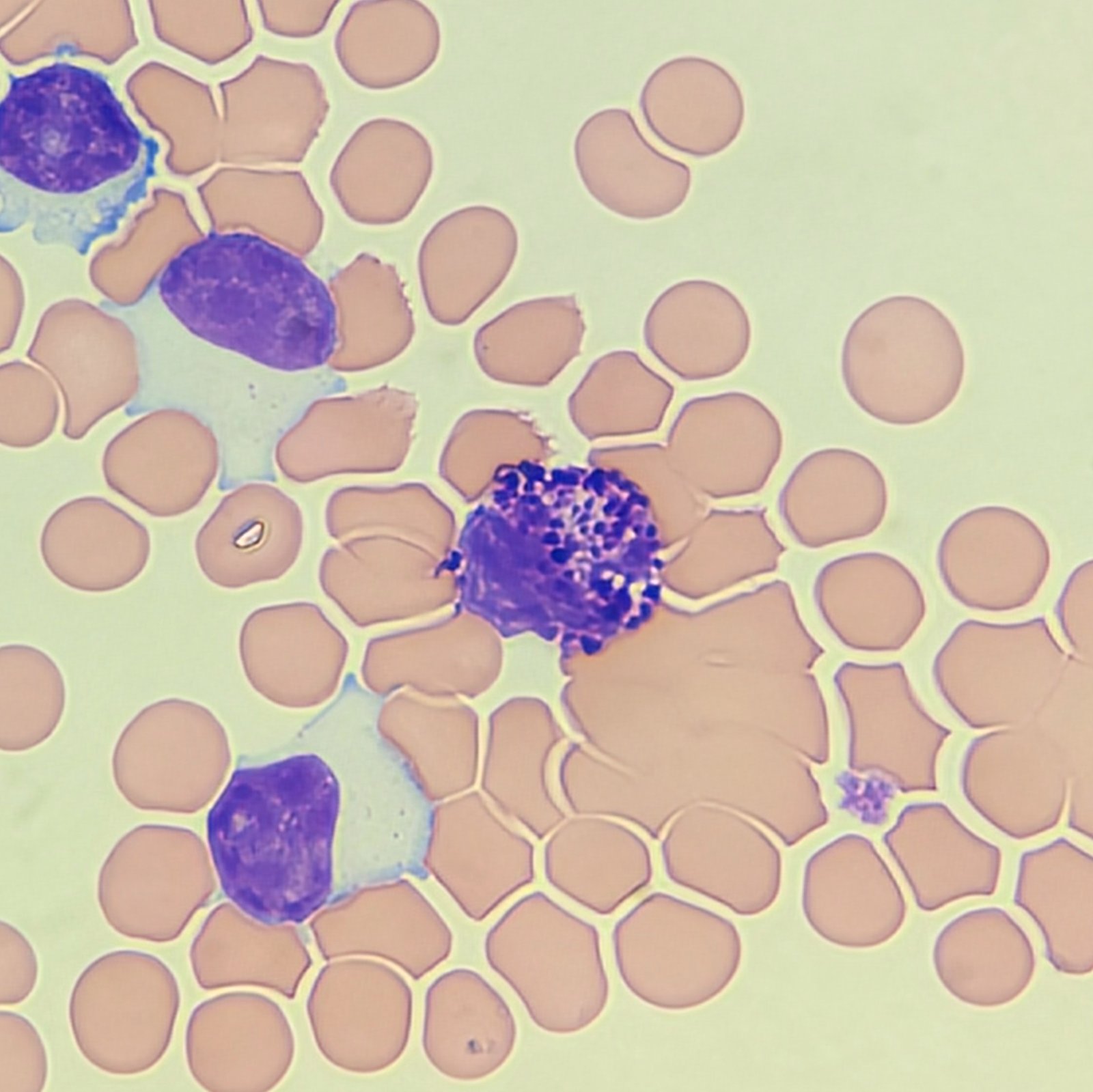
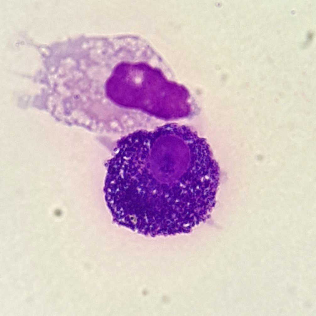
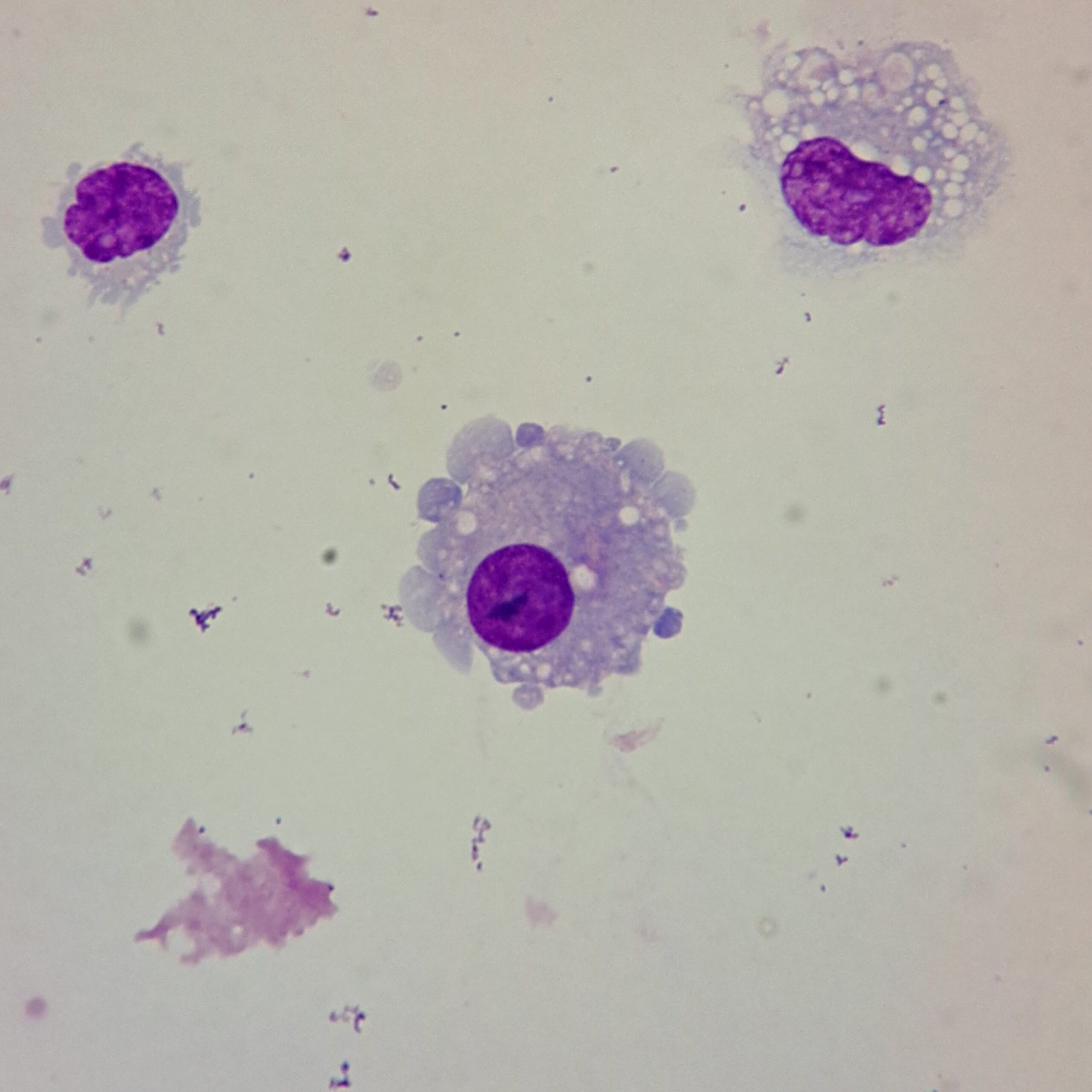
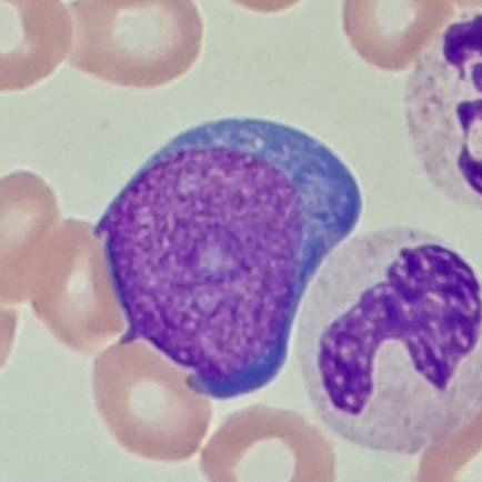
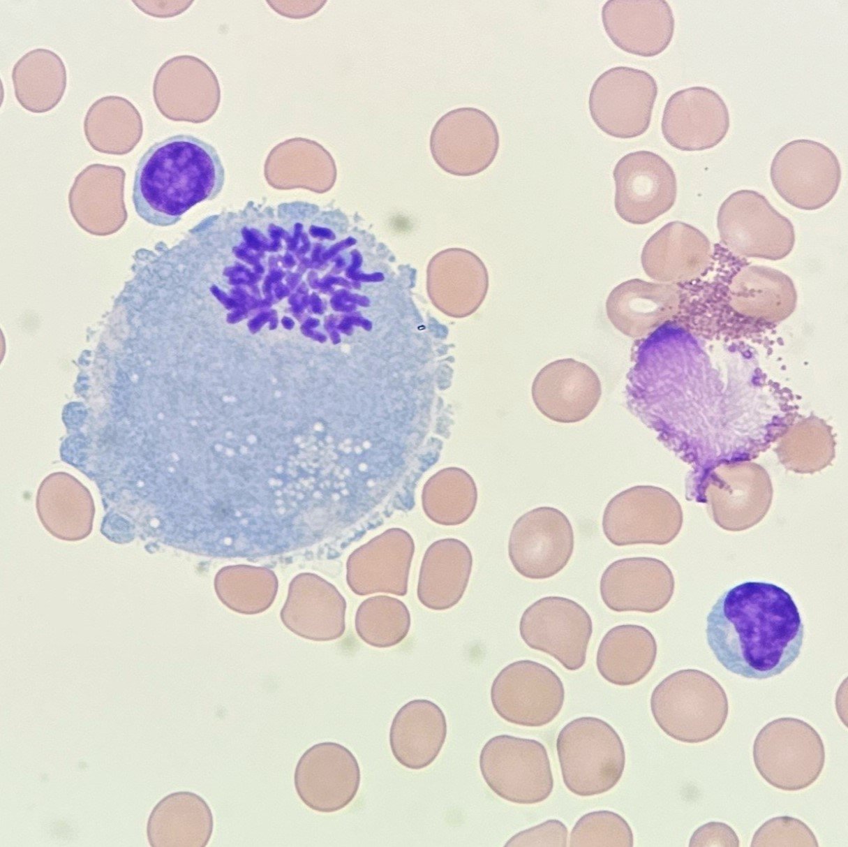
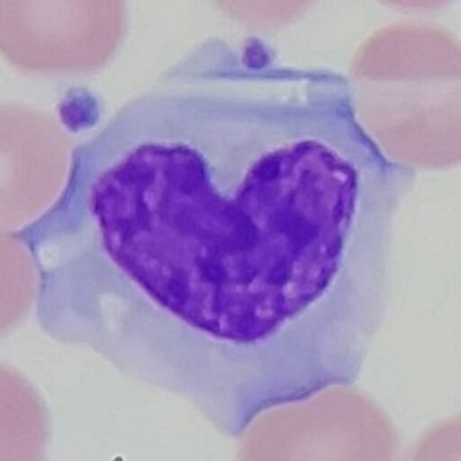
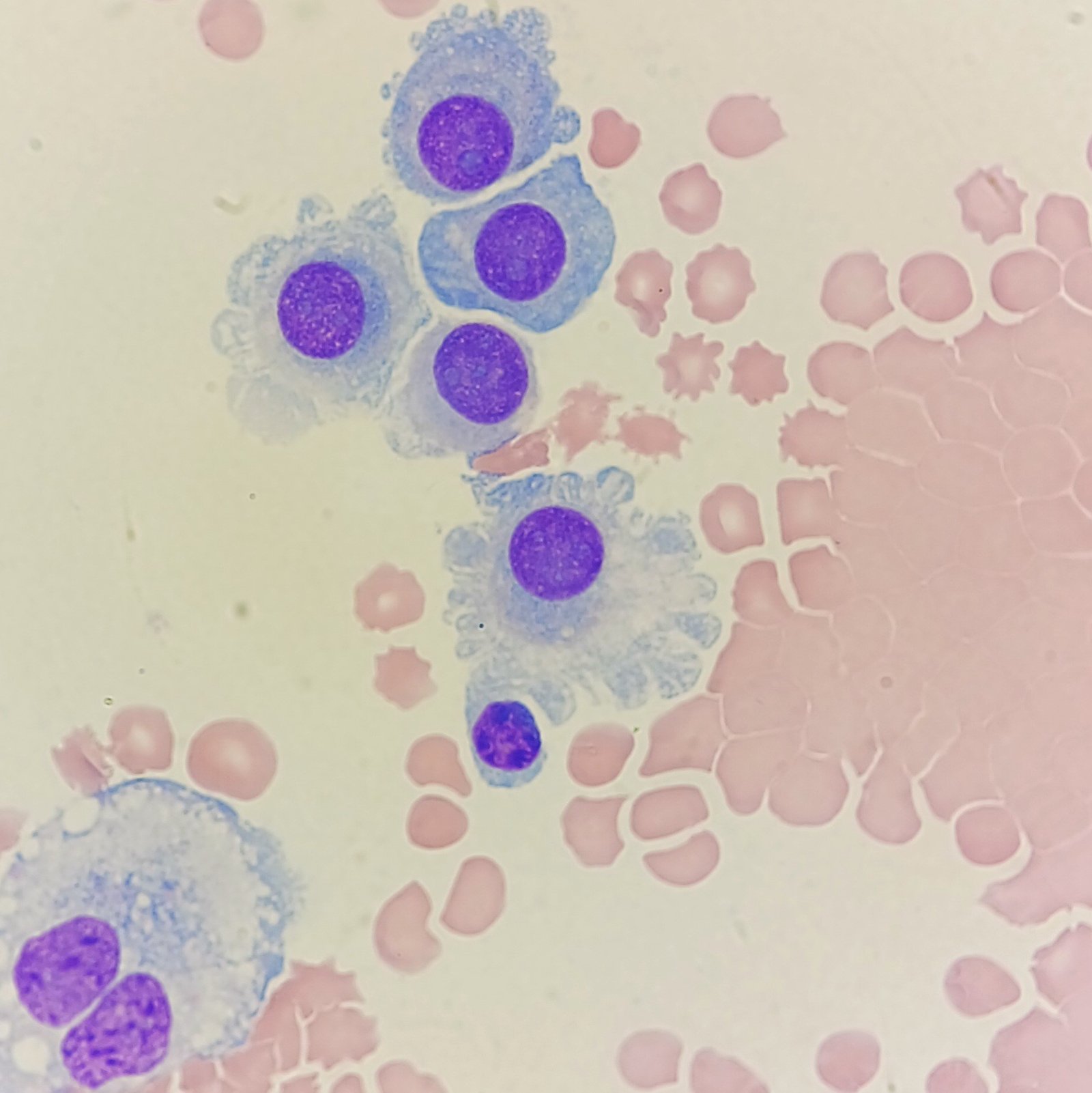
Mesothelial cells are described as having a “fried egg” appearance. They have a round to oval nucleus with smooth borders and evenly distributed chromatin. Nucleloli are usually present. Mesothelial cells may also be multinucleated.
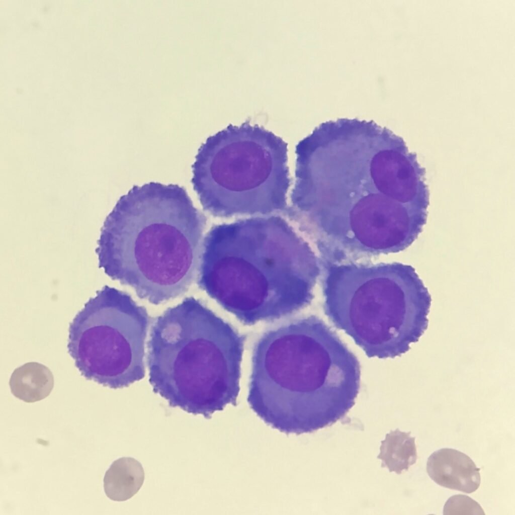
Cells may be seen in clumps, but “windows” between cells still allows for individual counting.
Lookalikes
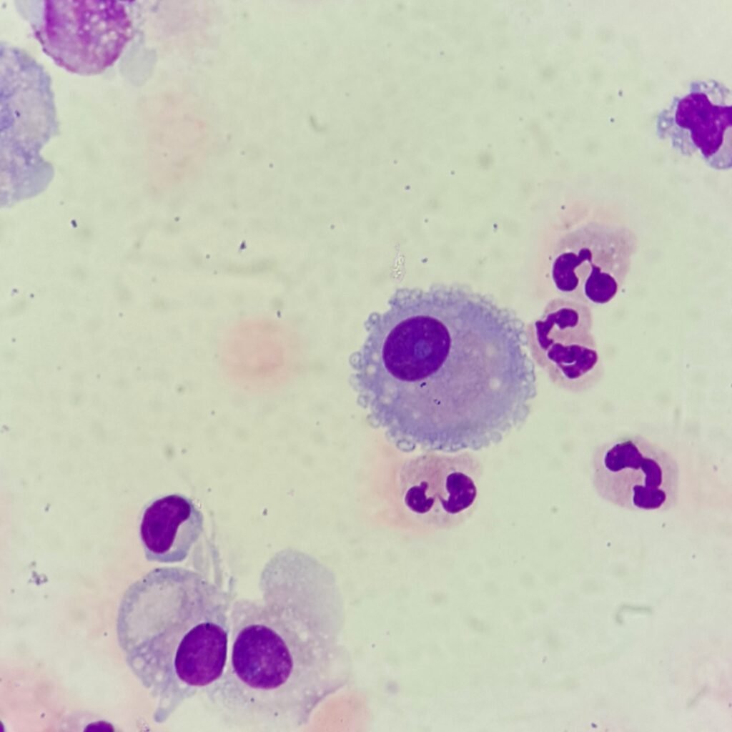
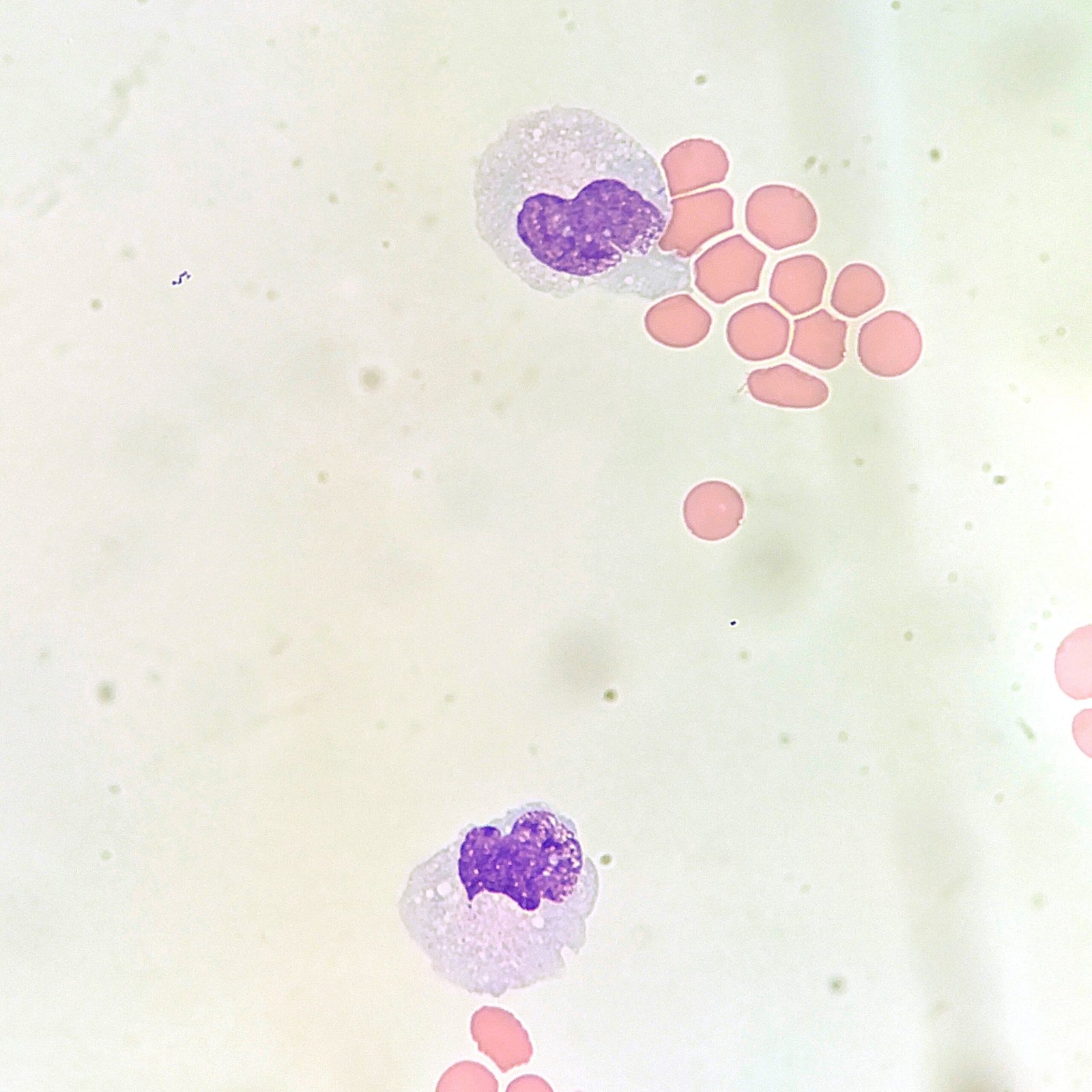
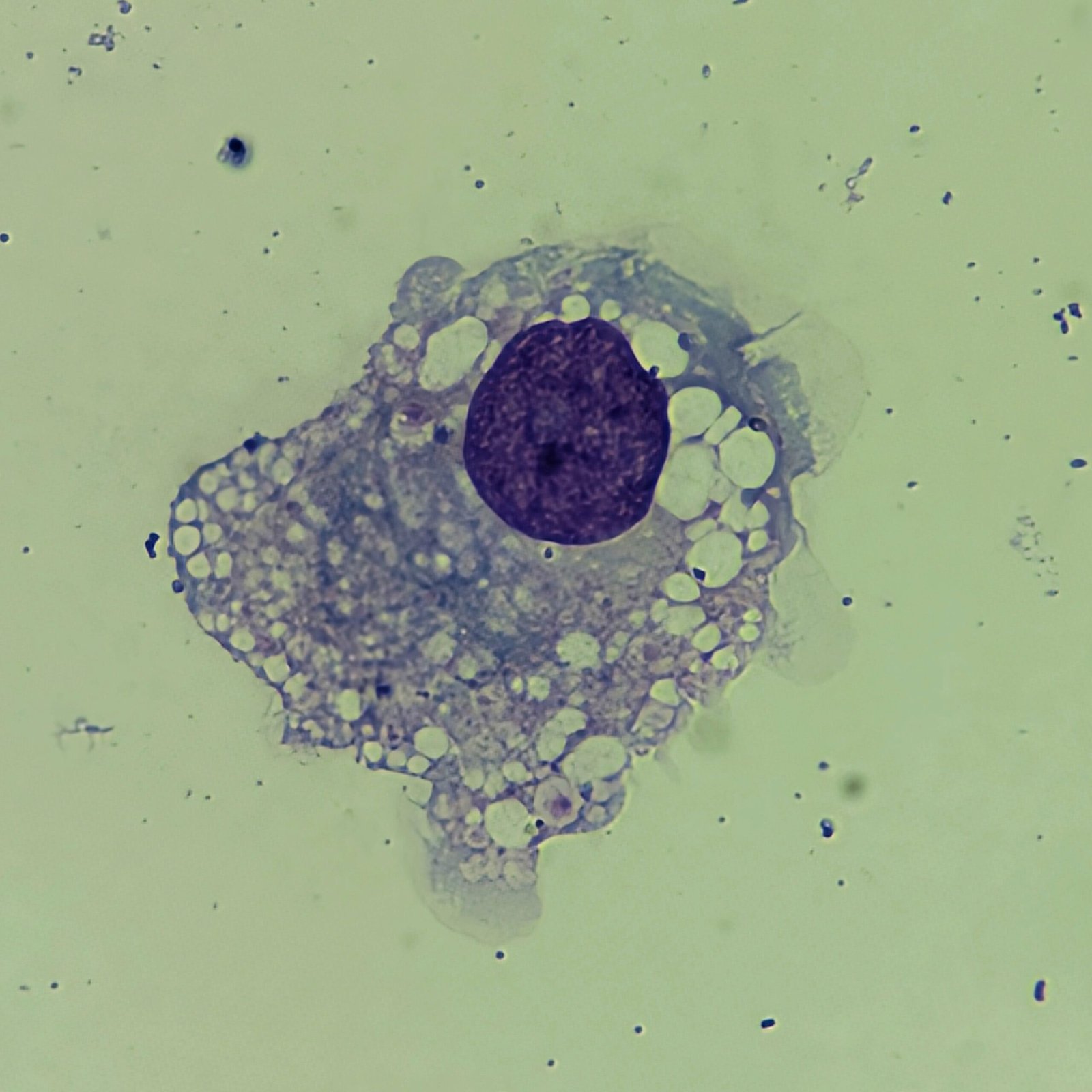
Macrophages are about the same size as mesothelial cells, so the two can often be confused. Macrophages can usually be differentiated by the presence of vacuoles and a lacey chromatin. If both cell types are present and differentiation is difficult, take a look around the slide to get an idea of each kind of morphology before starting a differential.
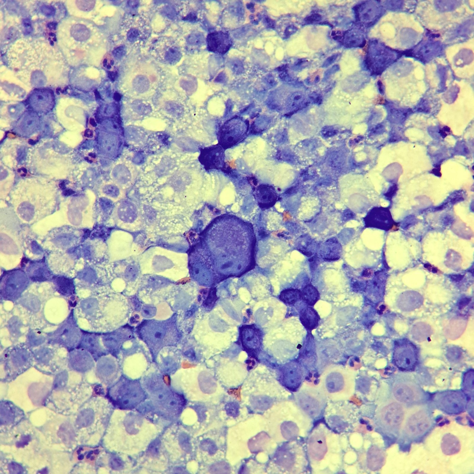
Both malignant cells and mesothelial cells can clump together. However, malignant cells will not have the windows between cells.
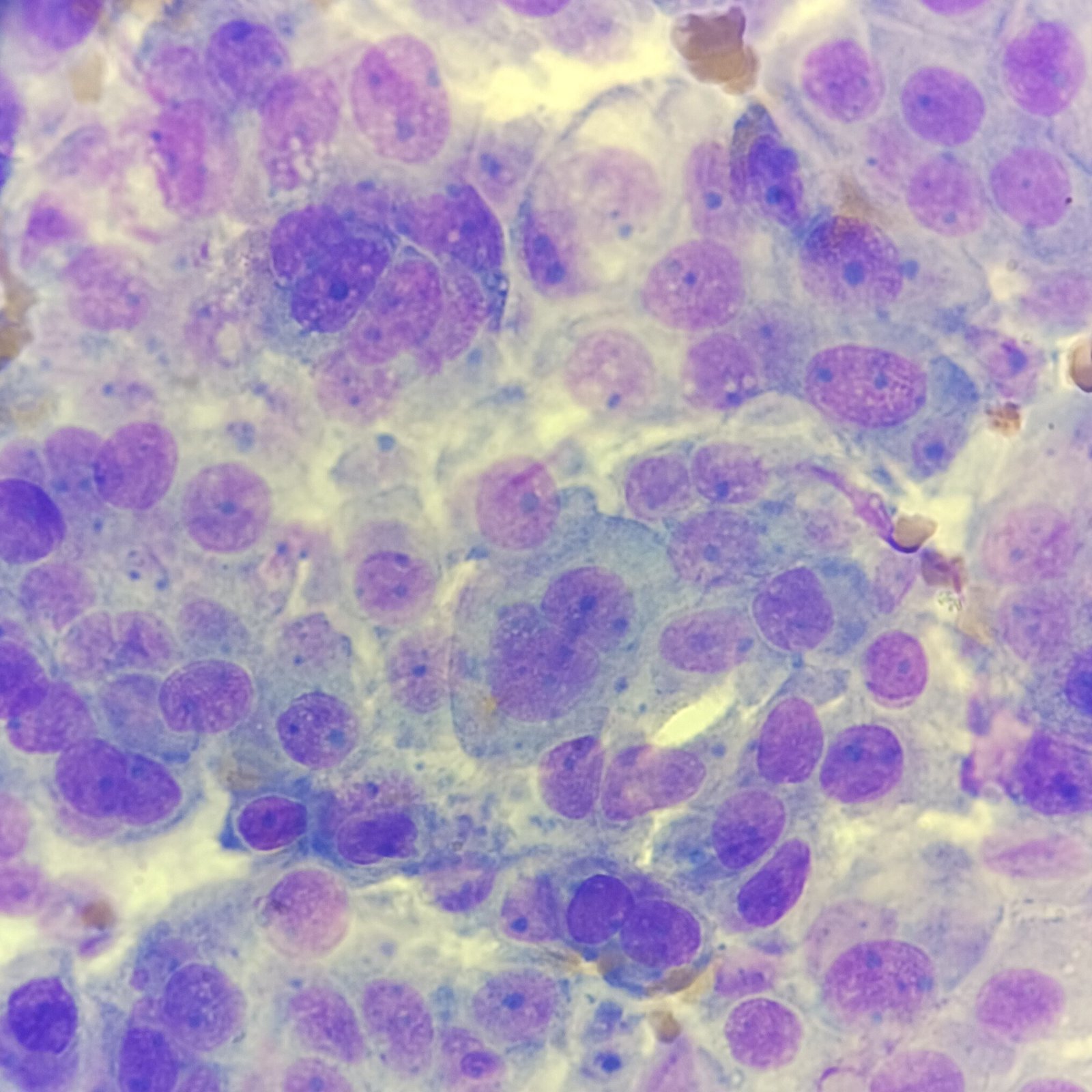
Gallery