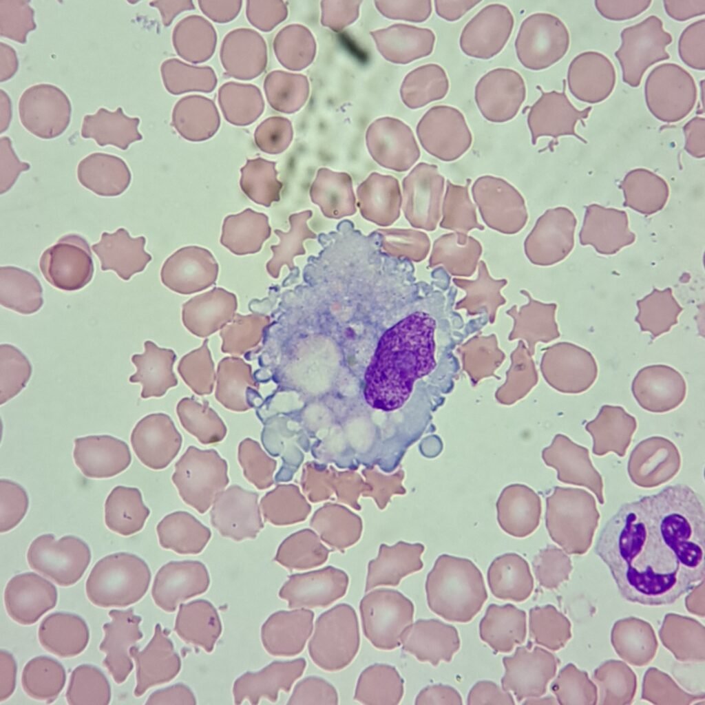
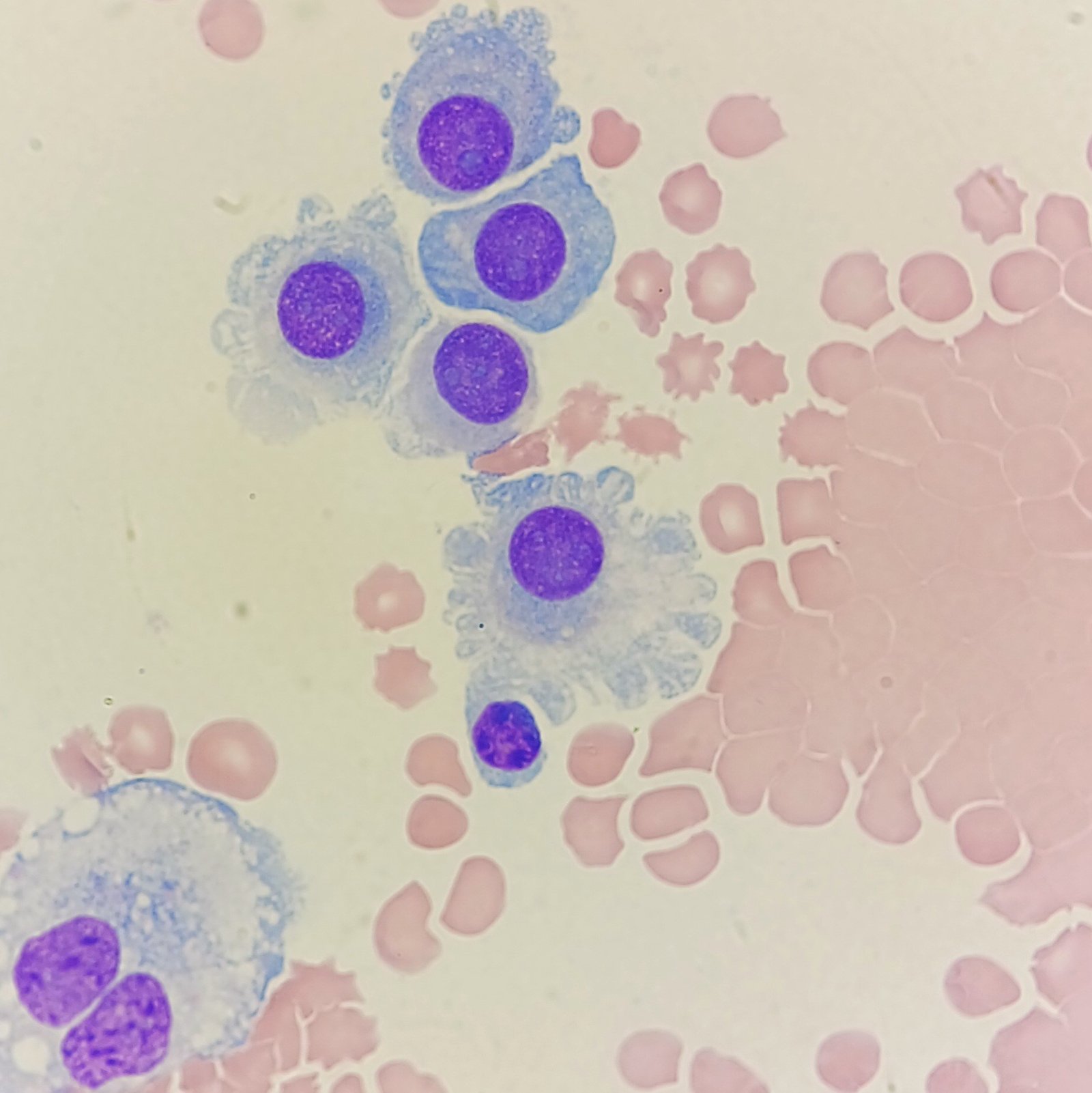
Monocytes in peripheral blood can migrate to tissue and differentiate into macrophages. Macrophages phagocytize debris or foreign material. Thus, they can be seen in body fluids ingesting bacteria, red blood cells, and even white blood cells at times. These cells may be increased with inflammation.
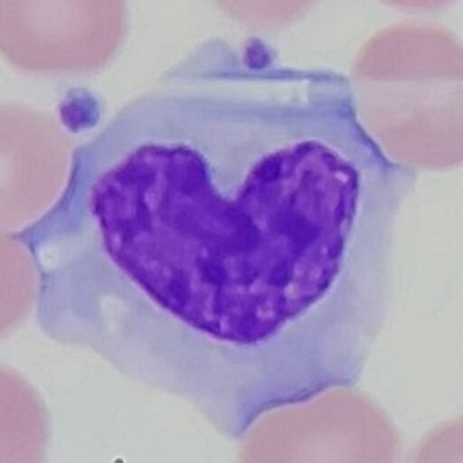
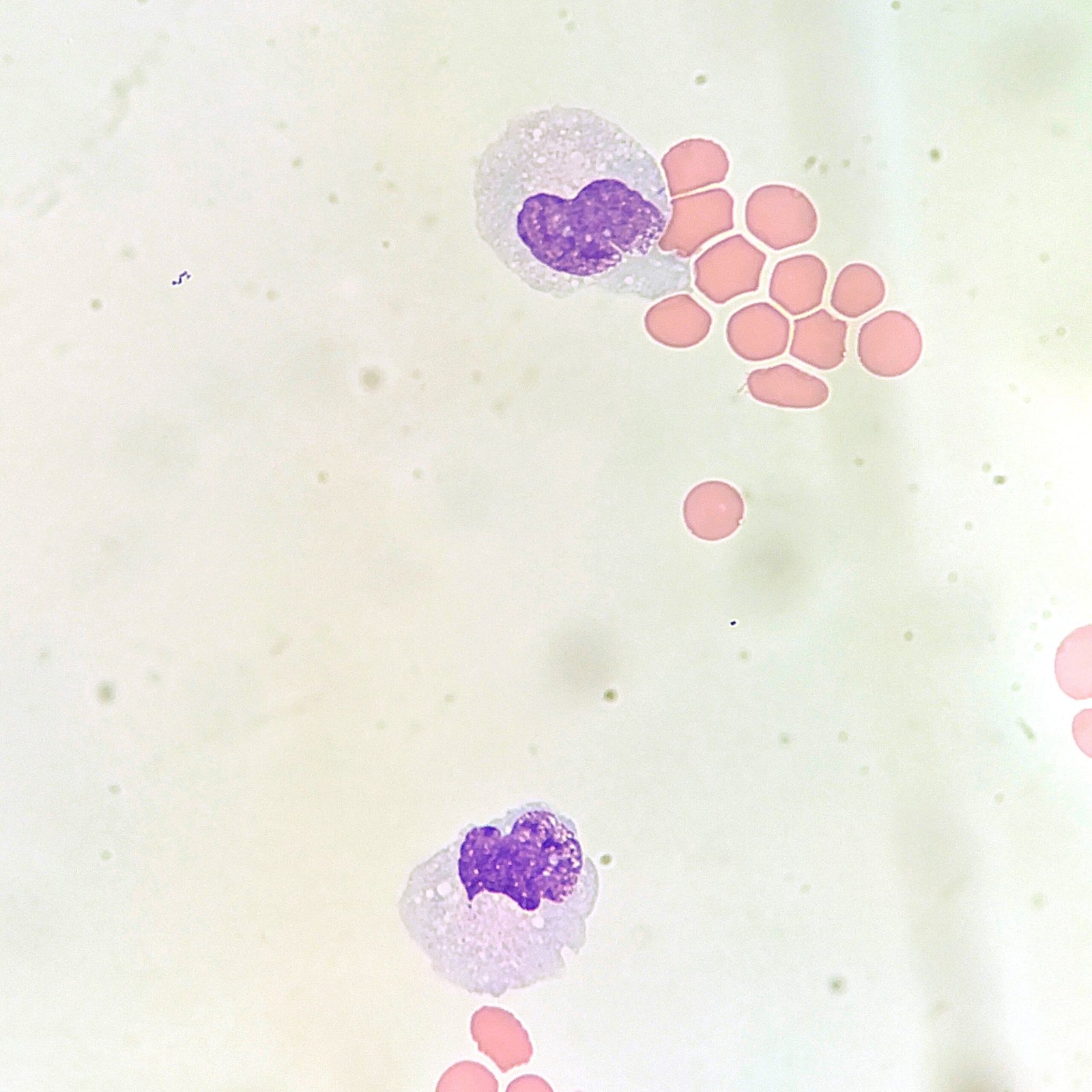
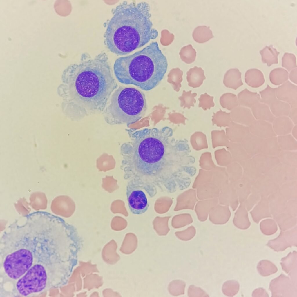
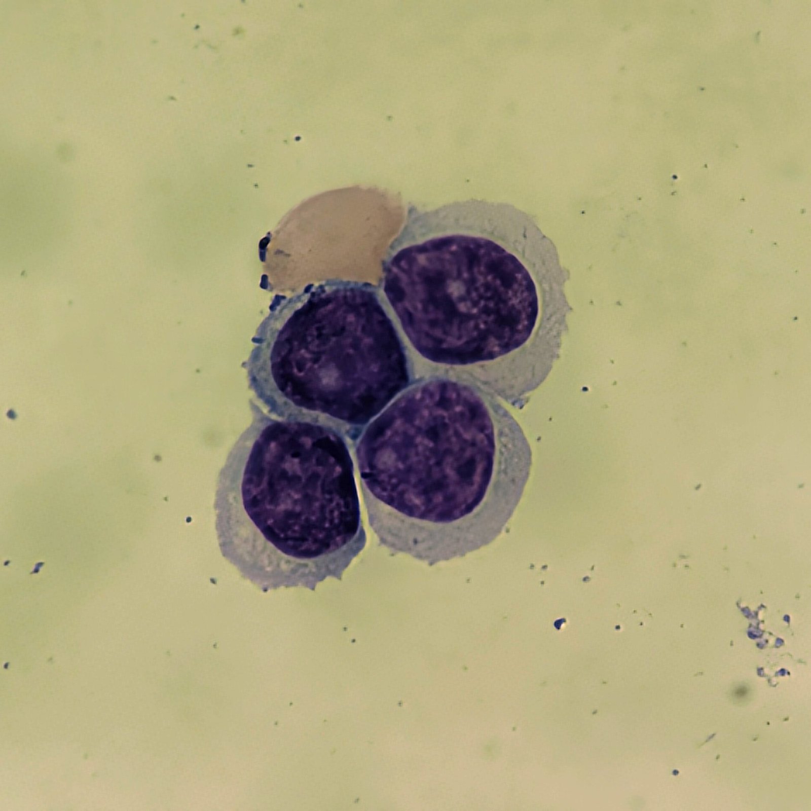
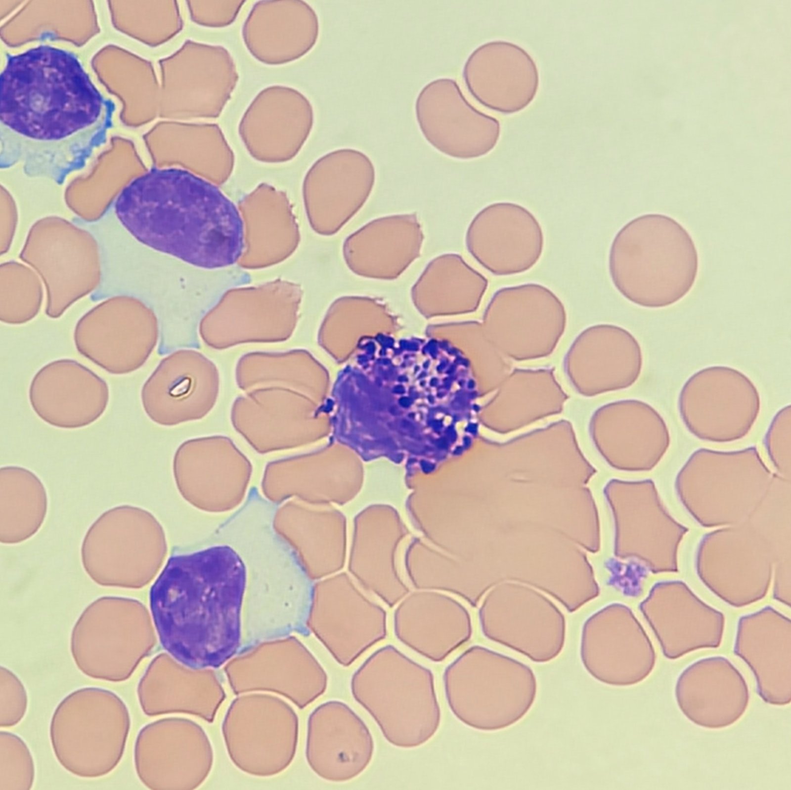
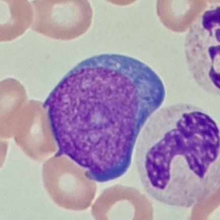
Monocytes
Monocytes in body fluids appear as they do in peripheral blood. Cytocentrifugation may exaggerate the vacuoles or make the cells appear larger.
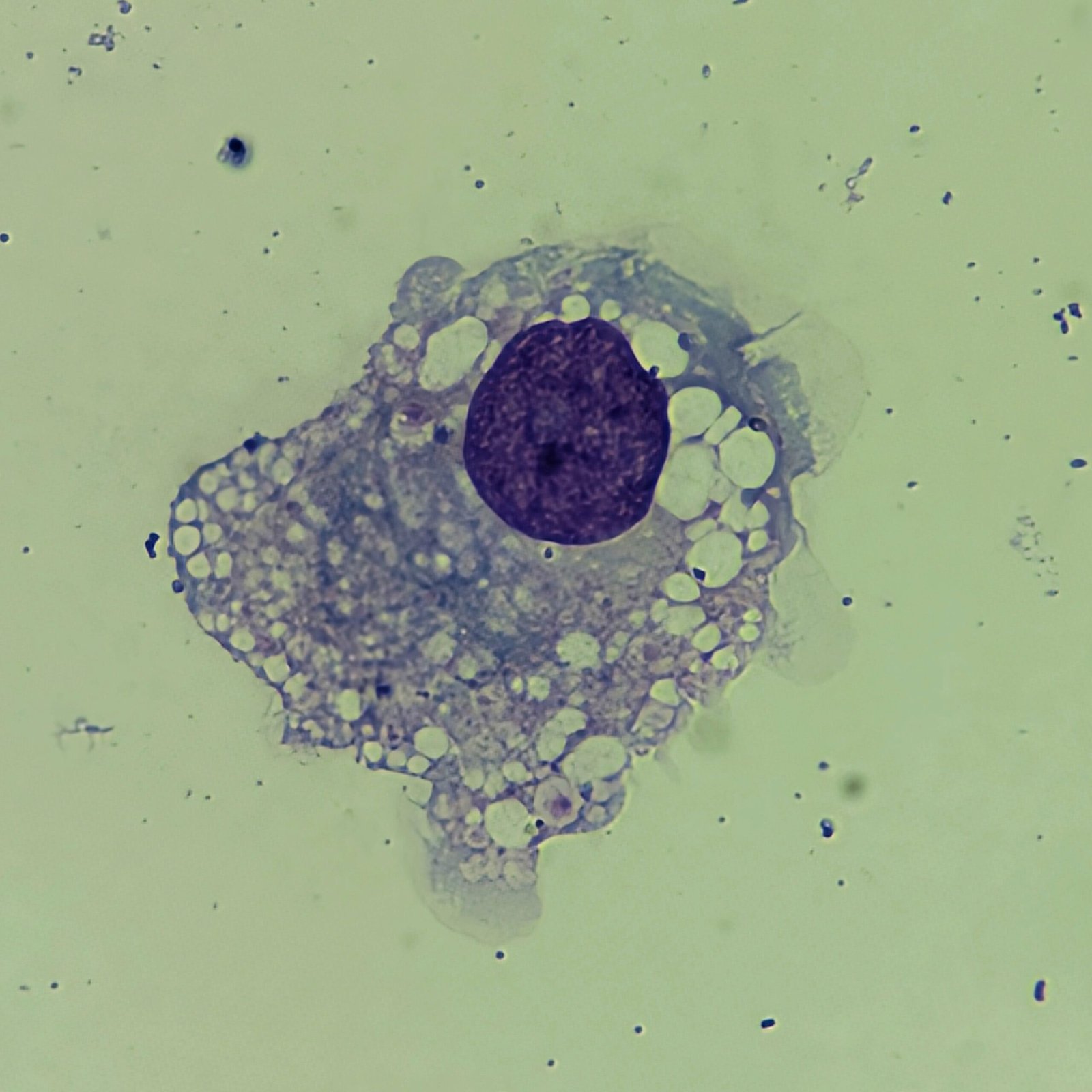
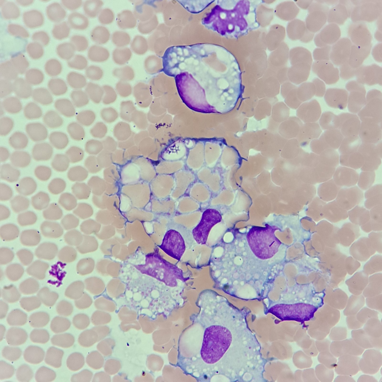
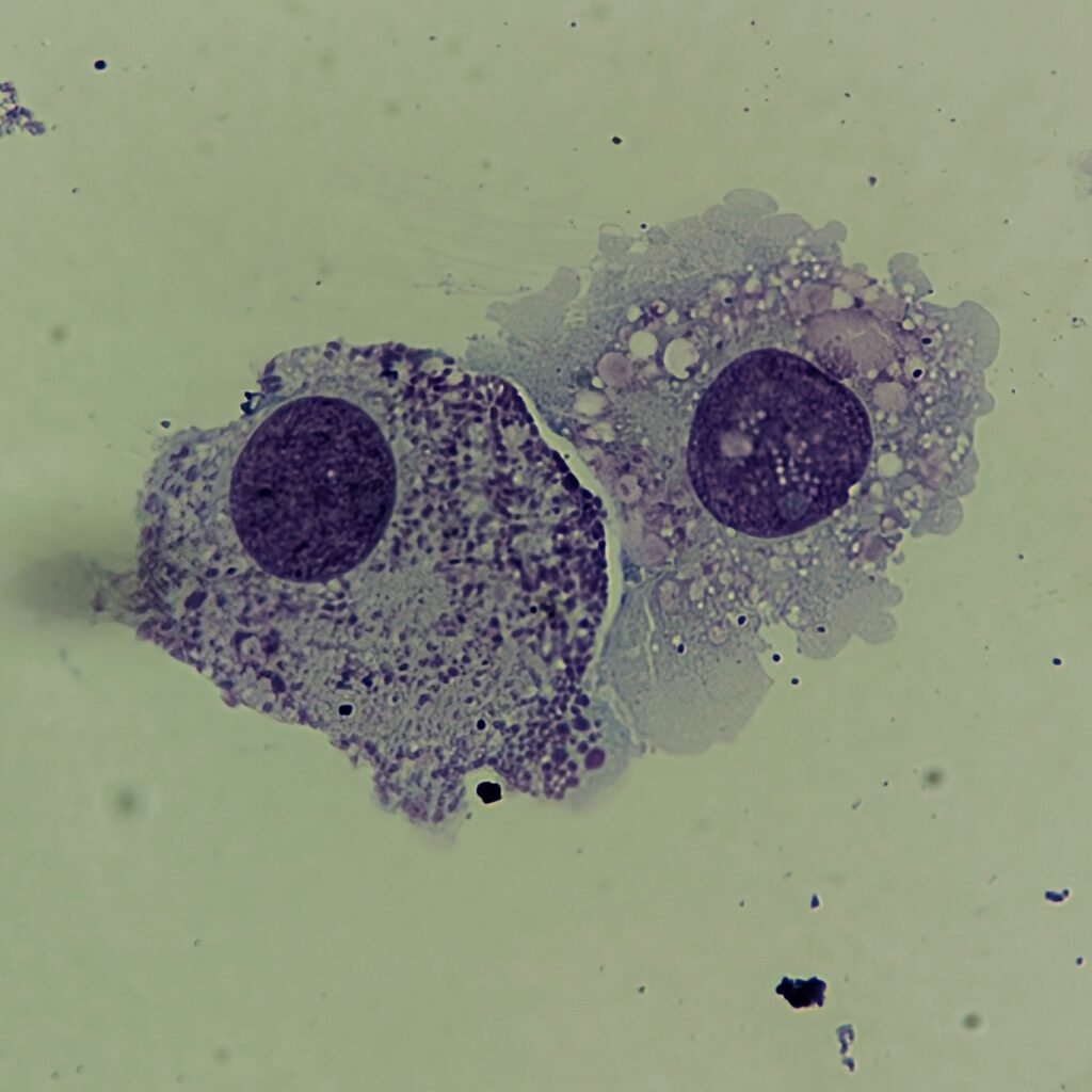
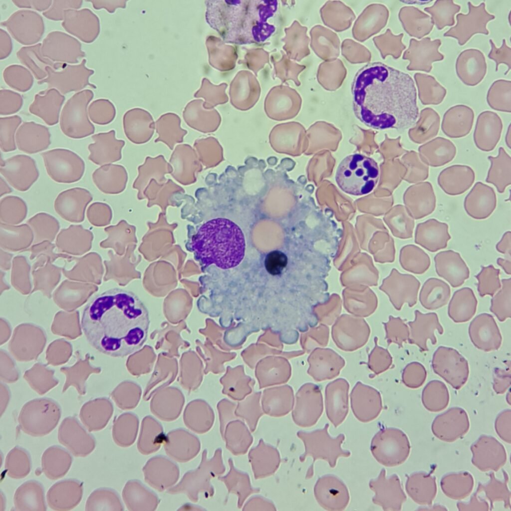
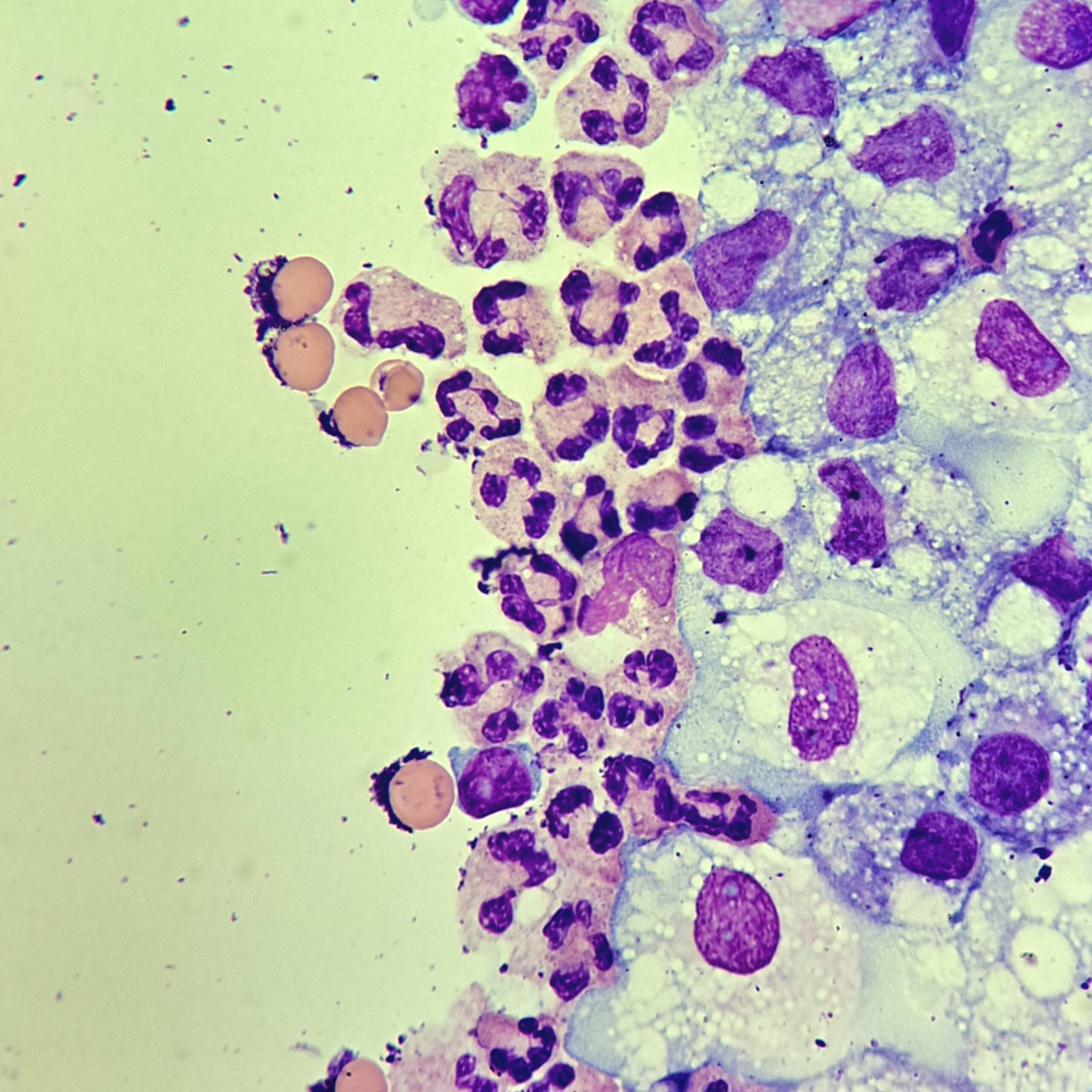
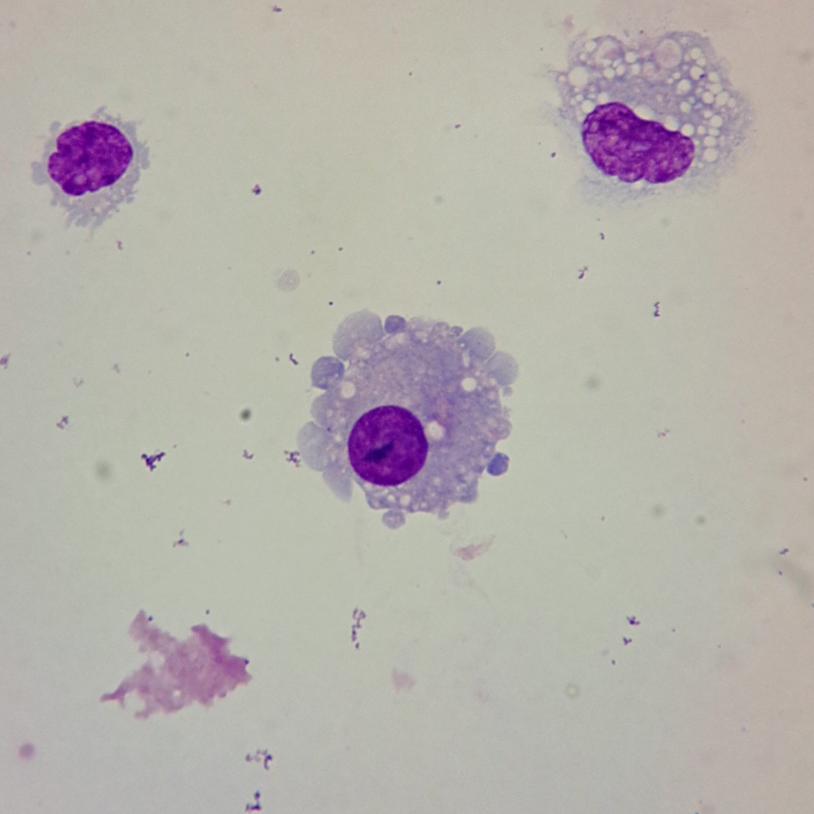
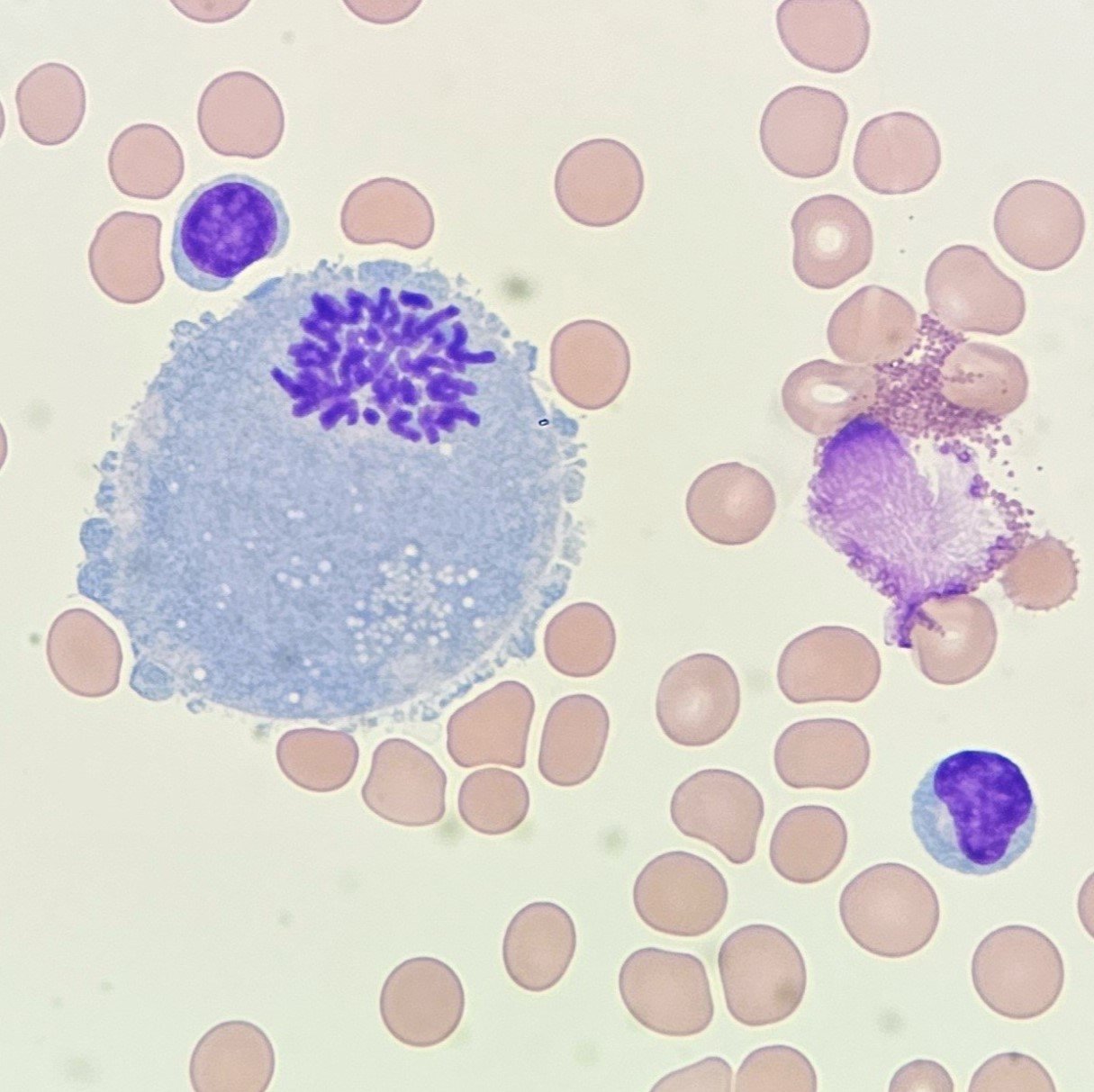
Macrophages
Macrophages are larger than monocytes and about the same size as mesothelial cells. Their cytoplasm often contains numerous vacuoles.
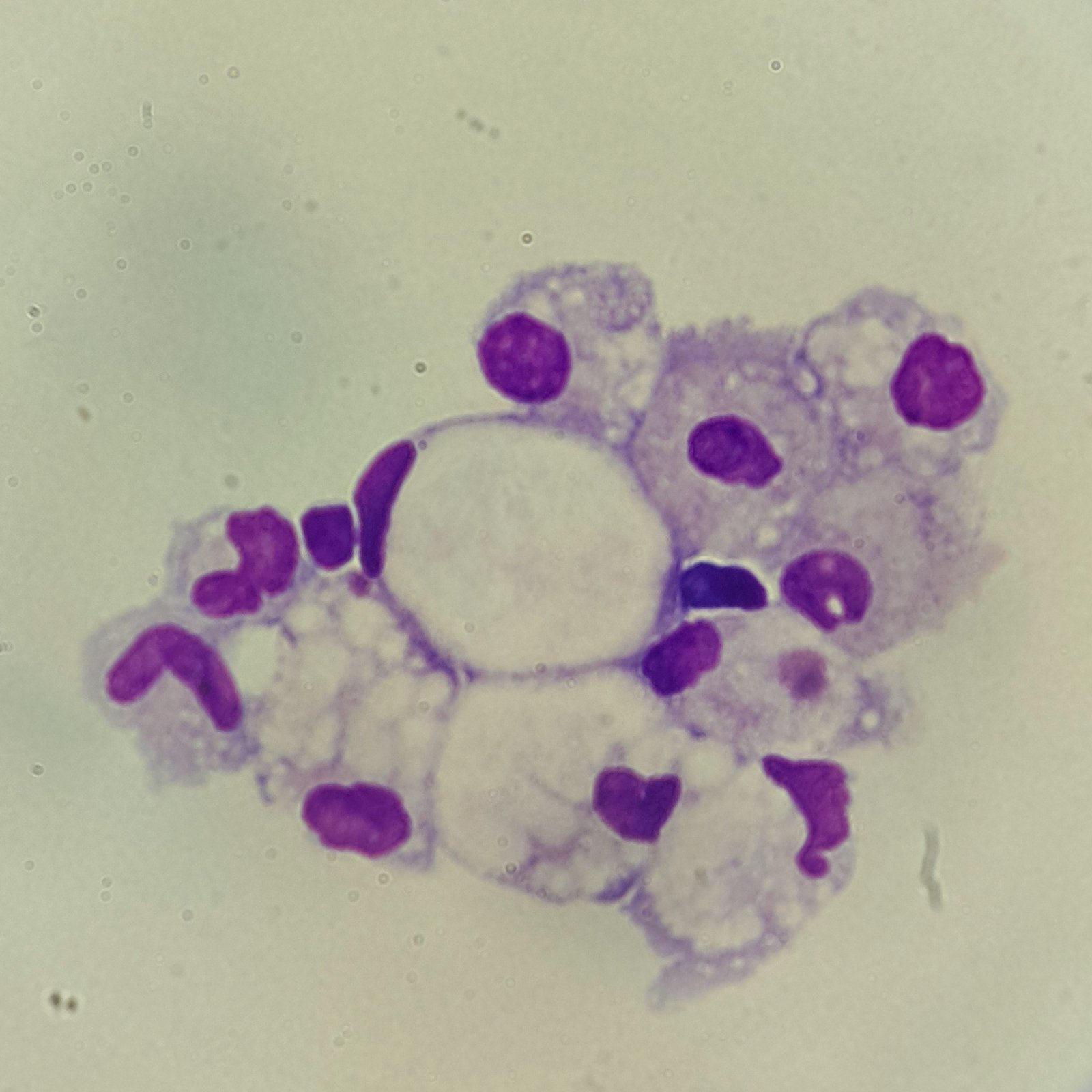
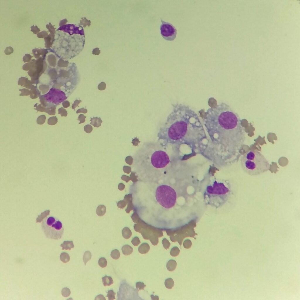
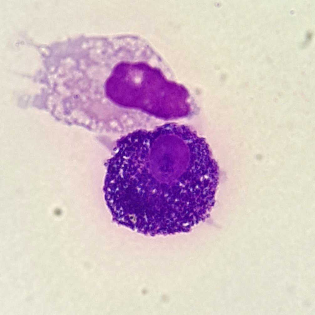
Erythrophages
Erythrophages are macrophages with engulfed red blood cells.
These cells can be seen after hemorrhage. They may also just be the result of the macrophage continuing phagocytic in vitro activity after collection.
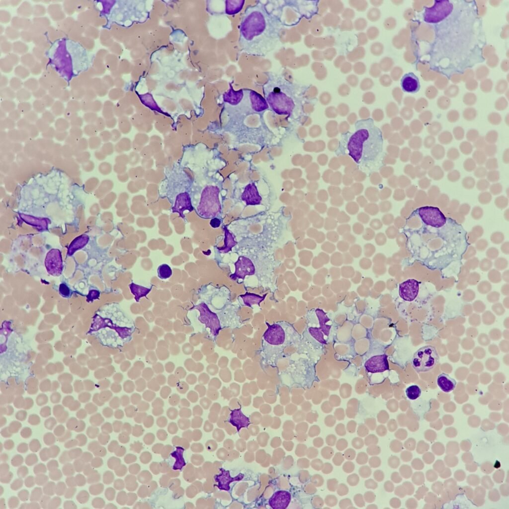
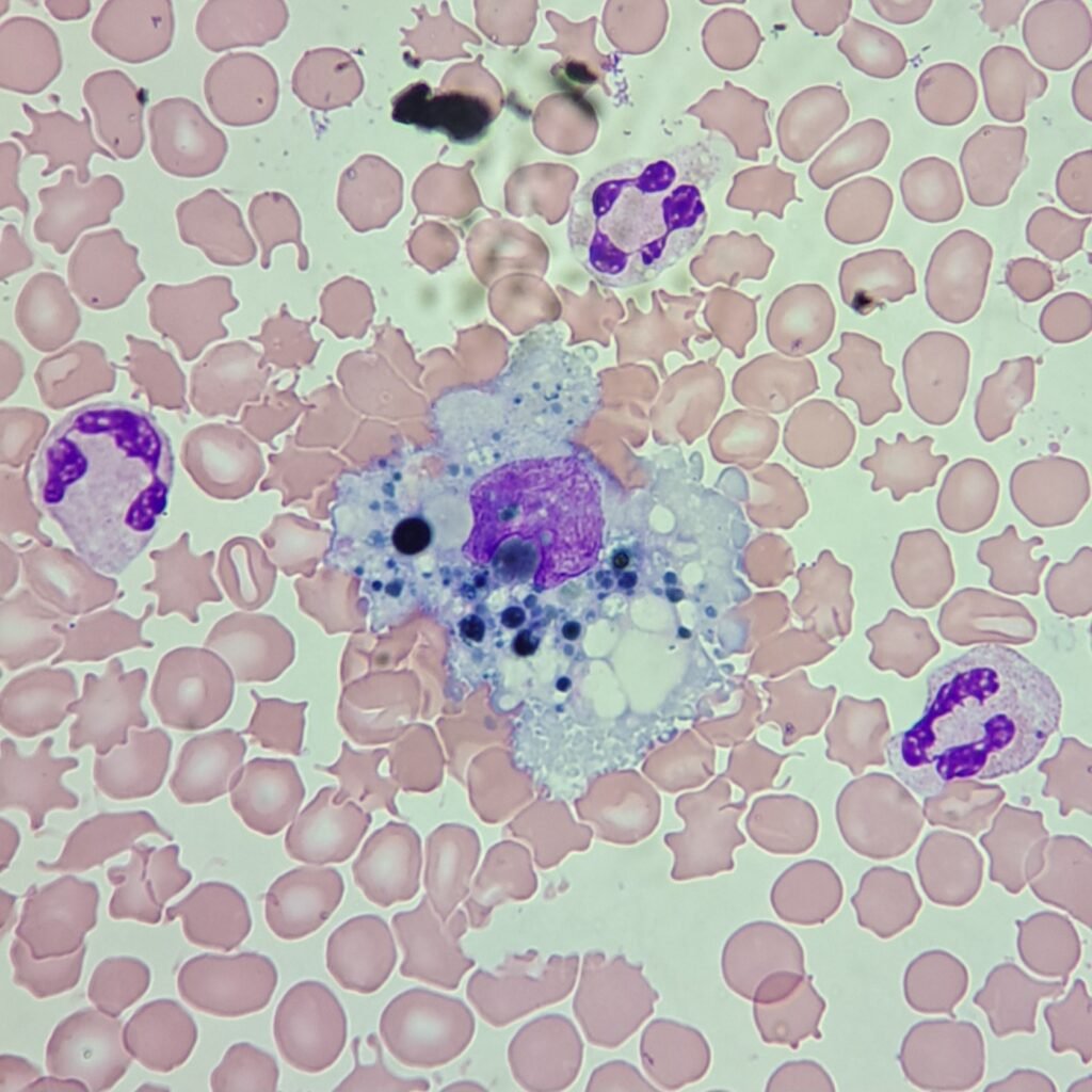
Lipophages
Lipophages are macrophages with abundant lipid vacuoles. These vacuoles may be full of extracellular lipids or the remnants of cell membranes. They may appear in CSF following a brain infarct.
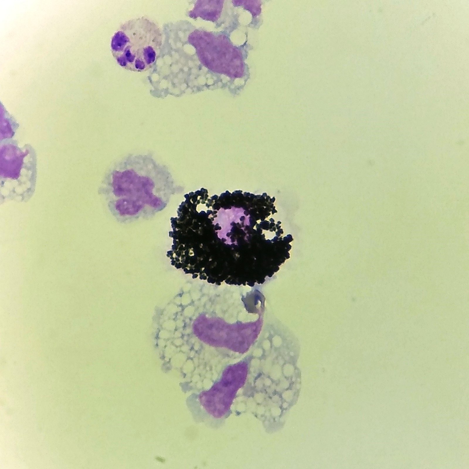
Siderophages
Siderophages are macrophages with coarse dark blue hemosiderin granules.
They can be seen even months after hemorrhage as macrophages phagocytose the hemoglobin remains of red blood cells.
They may also be seen in disorders where there is excessive iron buildup.
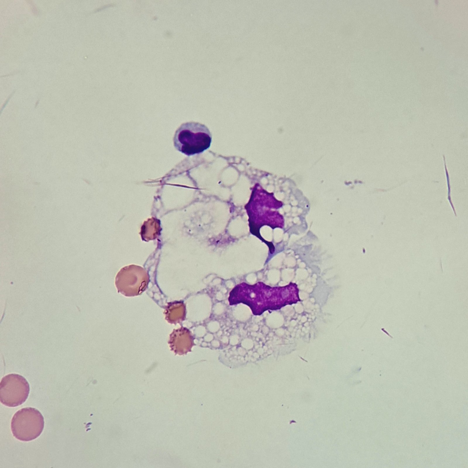
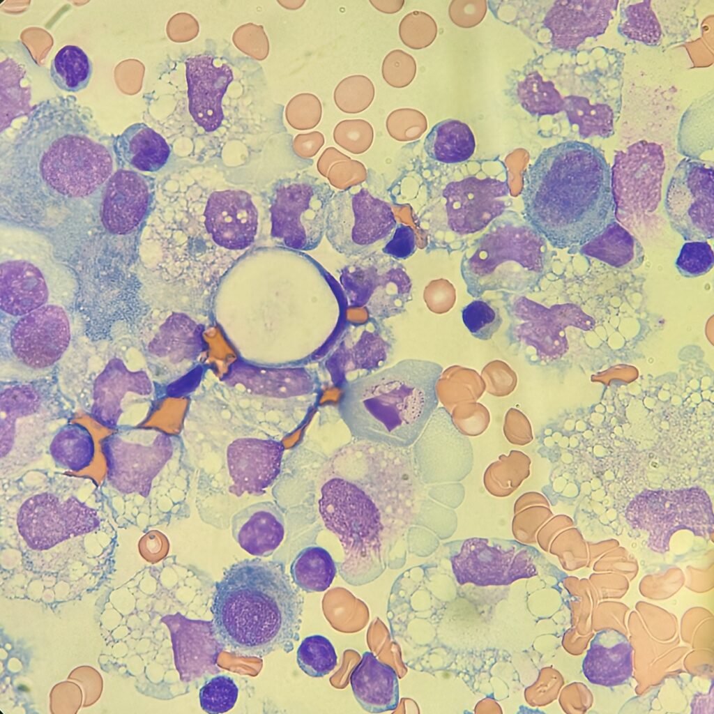
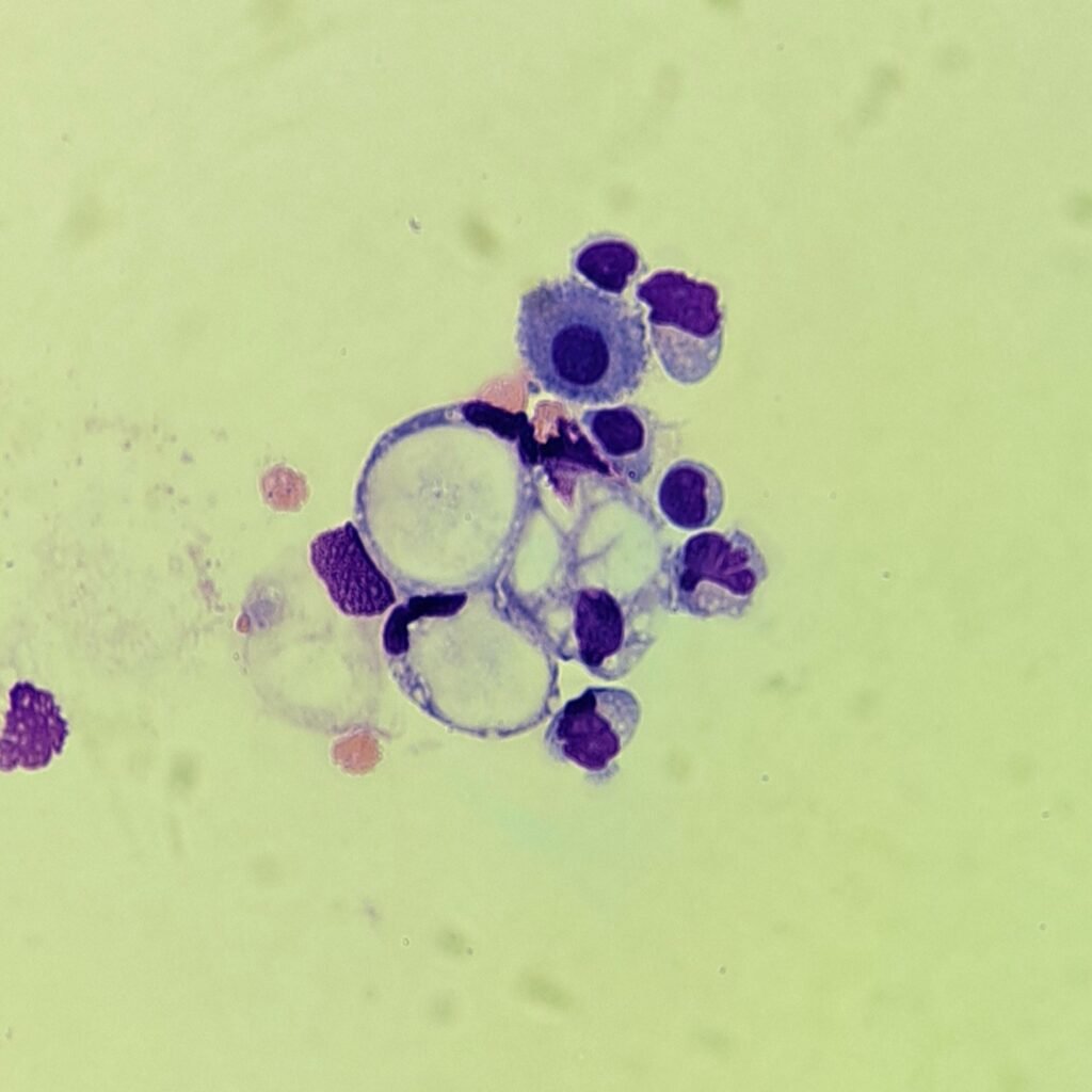
Signet Ring-Like
Macrophage has the appearance of a “signet ring” with several vacuoles forming what looks like the finger hole of a ring. The nucleus is pushed to one side of the cell, giving its distinctive appearance. Note that this type of macrophage is different from a true signet ring cell.
May also be seen in disorders where there is excessive iron buildup.
Lookalikes
Mesothelial cells have a wide variety of morphologies. They are about the same size as macrophages, so they can be confusing. Macrophages can usually be differentiated by the presence of vacuoles and a lacey chromatin. If both cell types are present and differentiation is difficult, take a look around the slide to get an idea of each kind of morphology before starting a differential.
Lymphocytes in body fluids appear larger due to cytocentrifugation and can look similar to monocytes. However, monocytes have a ground-glass cytoplasm that often has vacuoles. The nucleus has lacey chromatin and is often indented, as opposed to lymphocytes which have clumpy chromatin and a round nucleus.

Signet ring-like macrophages can be mistaken for true signet ring cells.
This article on cytojournal.com has more information on differentiation.

Gallery
Other Body Fluid Cells

Additional Resources
- Monocyte and macrophage differentiation: circulation inflammatory monocyte as biomarker for inflammatory diseases – PMC
- Monocytes and Macrophages: Macrophage and Monocyte Function, Origin and Related Conditions | Technology Networks
- Macrophages – Definition, Function, vs Monocytes, vs Neutrophils etc.
- Monocyte/Macrophage, % – Cell Count and Differential, Synovial Fluid – Lab Results explained | HealthMatters.io
- BFBRG_BodyFluidBench_Sample.pdf
- Indicators of true intracerebral hemorrhage: hematoidin, siderophage, and erythrophage
- Signet-ring cells in pleural and peritoneal effusions identified on Wright stains – A diagnostic pitfall – CytoJournal
- Macrophage in BALF
- 10: Pleural, Pericardial, and Peritoneal Fluid Analysis