Synoviocytes are lining cells seen in synovial fluid. They are not clinically significant.
Appearance
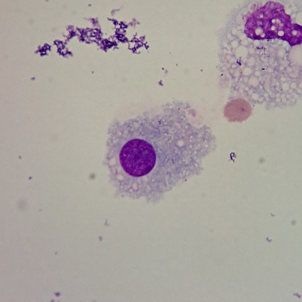
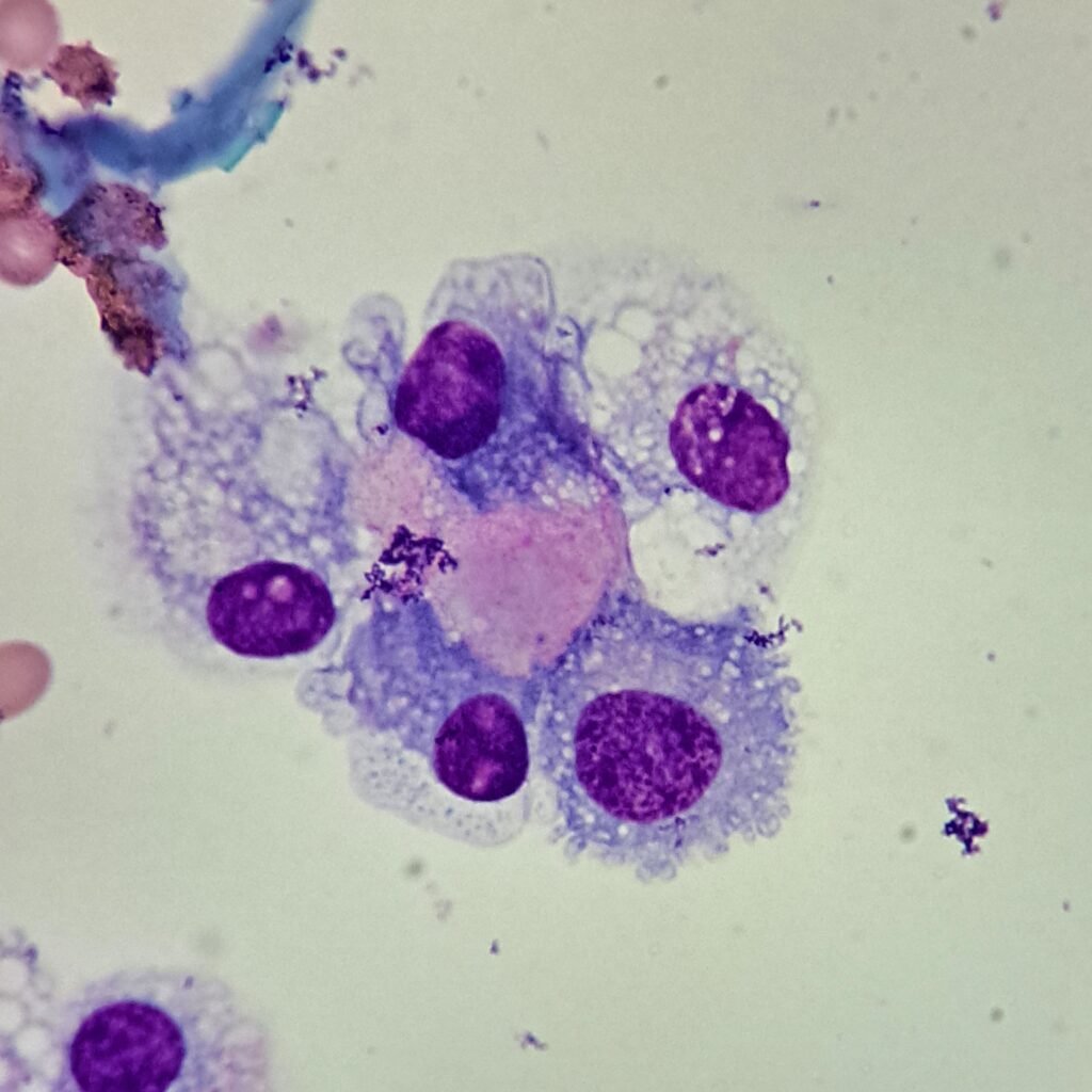
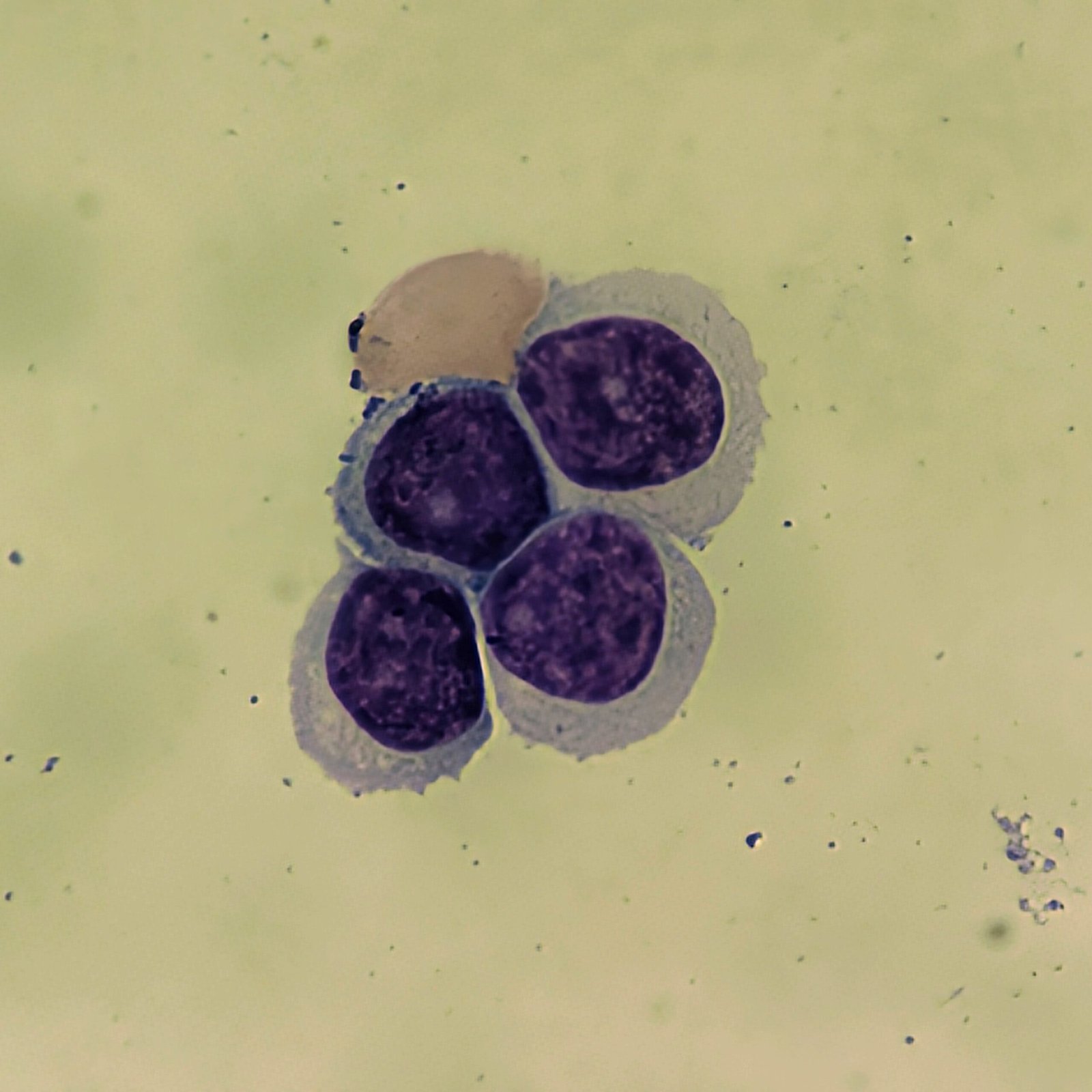
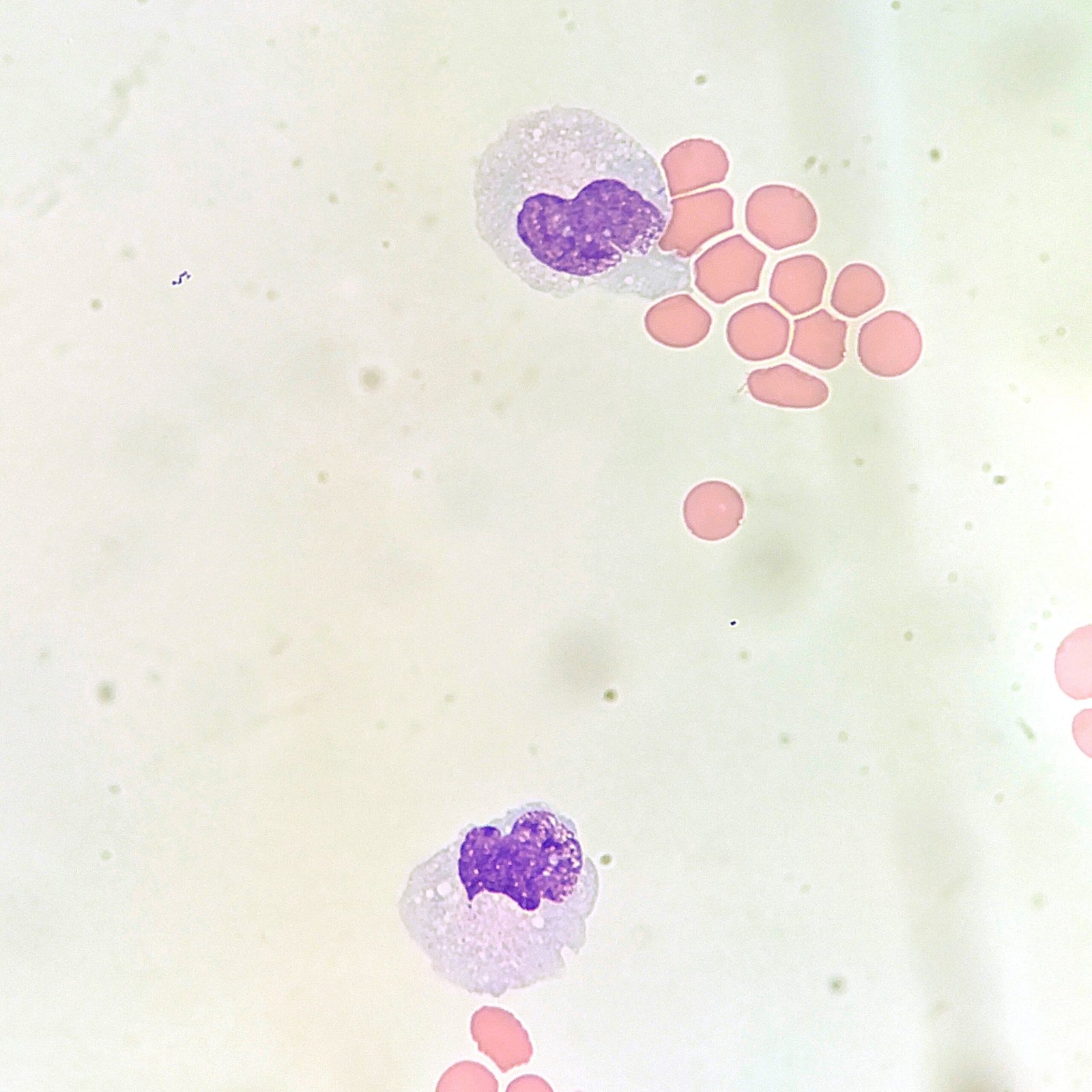
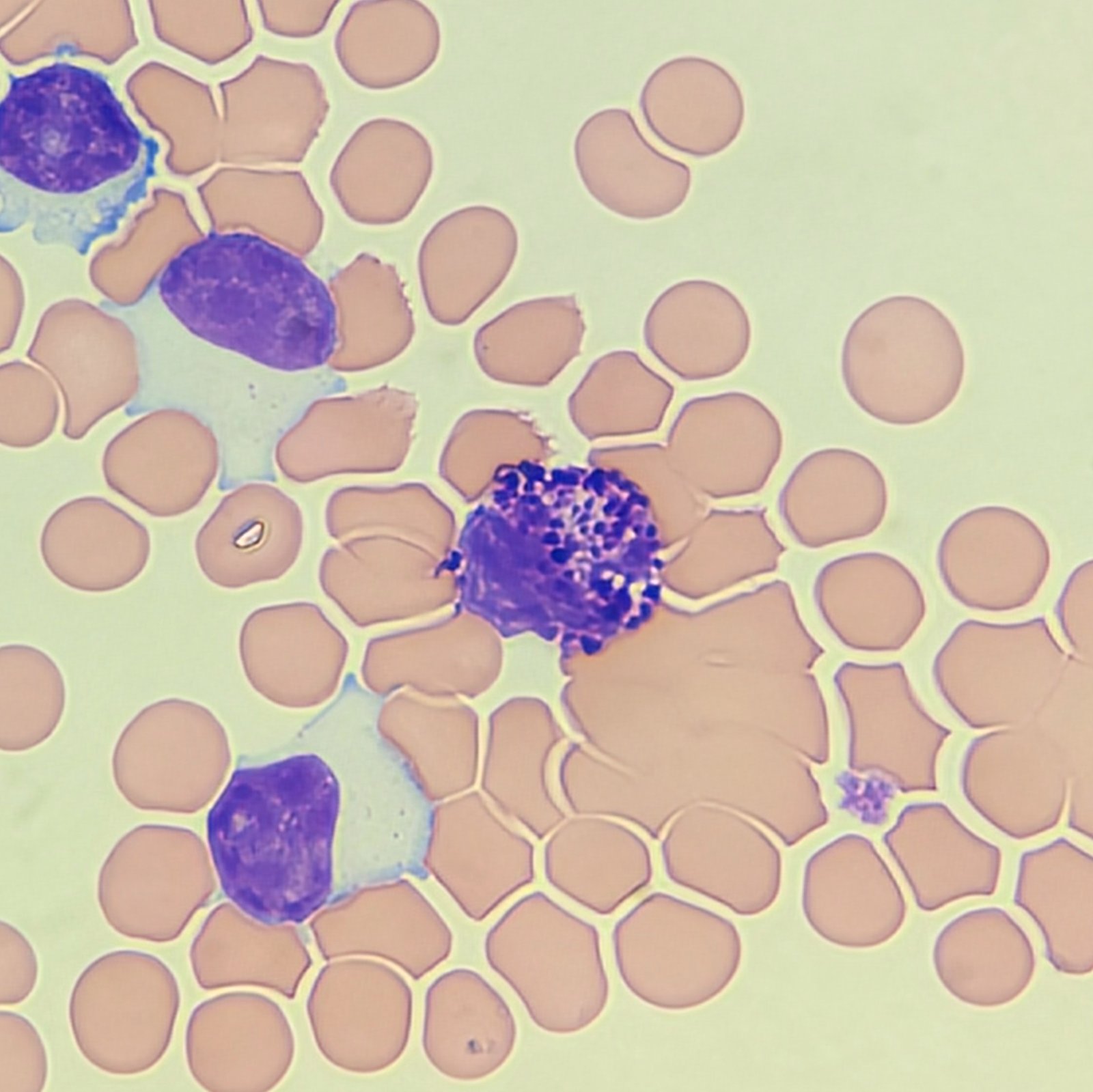
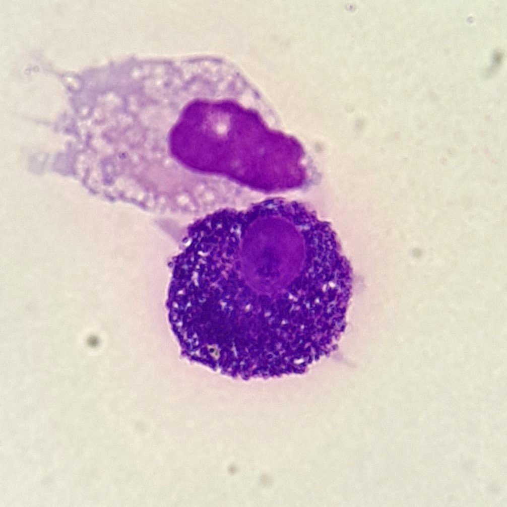
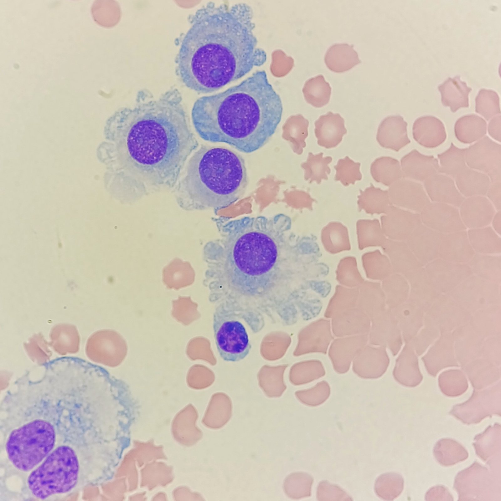
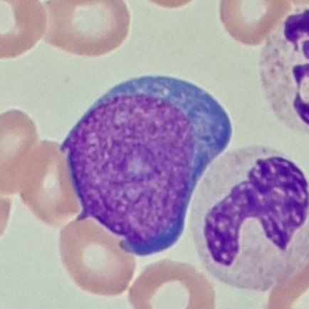
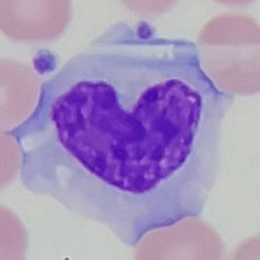
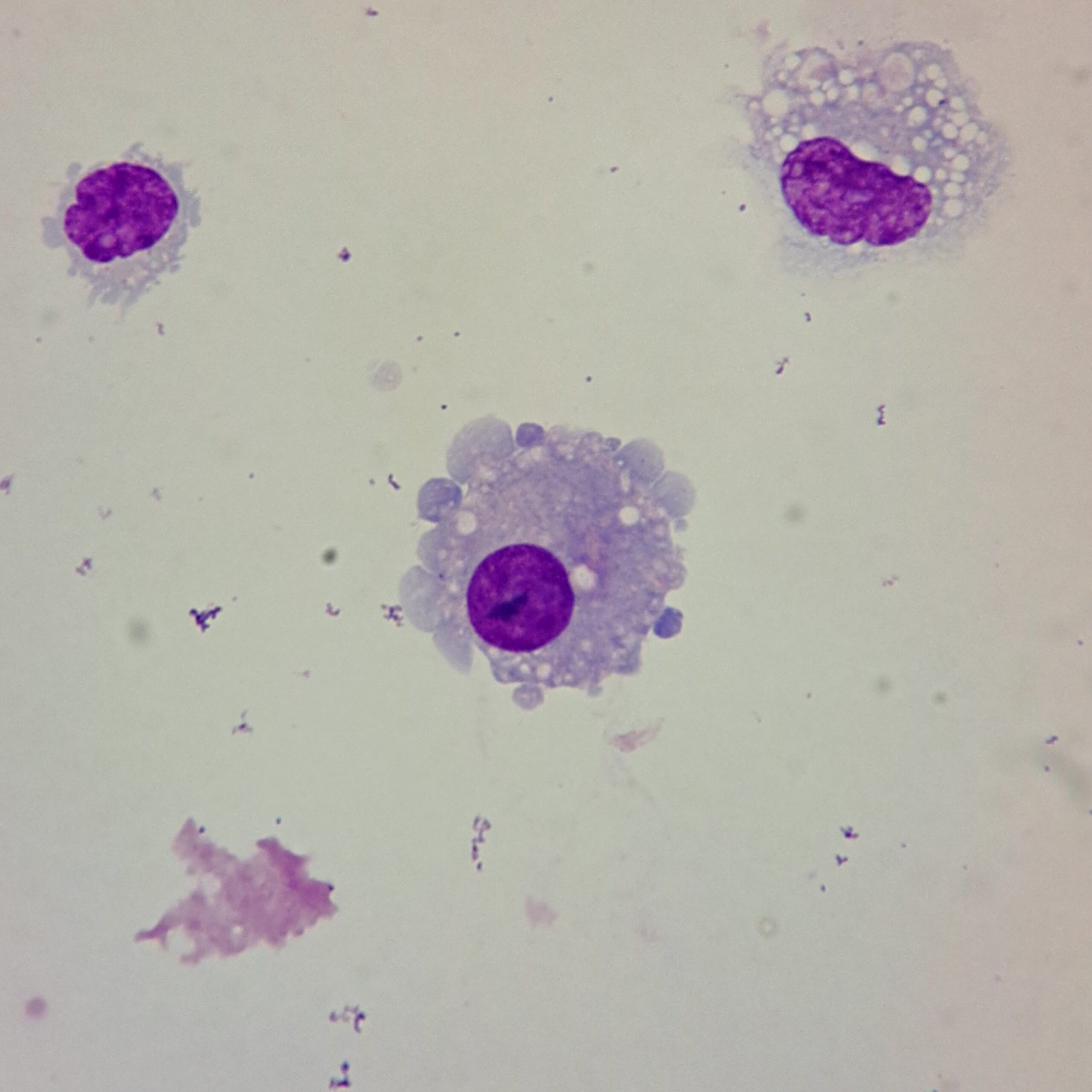
Synoviocytes are visually similar to mesothelial cells. The nucleus is round to oval with smooth borders. Nucleoli may be present. Cytoplasm may have irregular edges or vacuoles.
Lookalikes
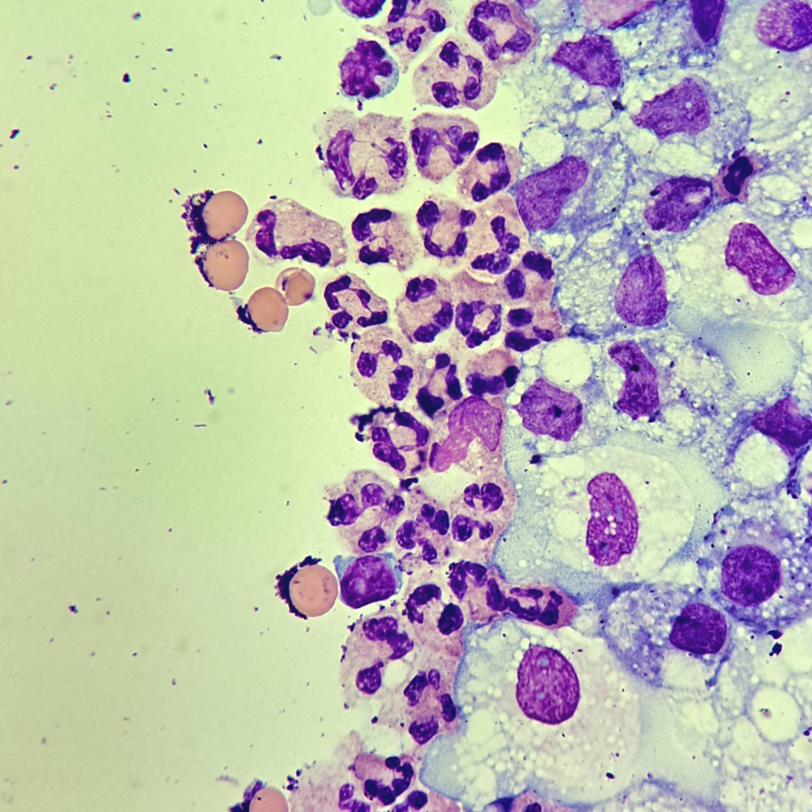
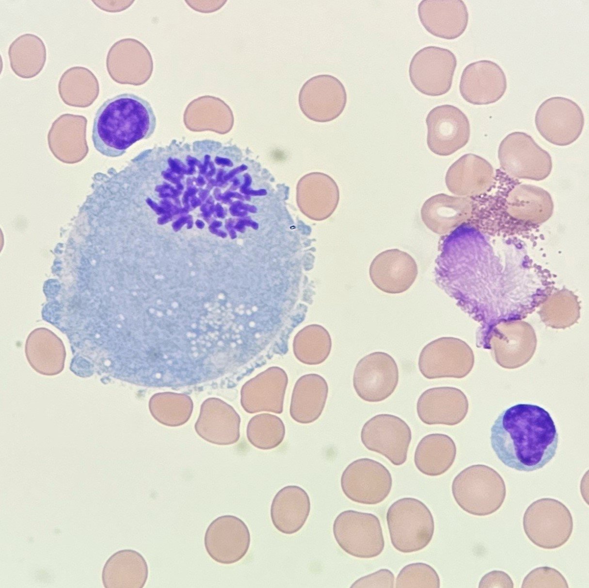
Macrophages are about the same size as synoviocytes, so the two can often be confused. Macrophages can usually be differentiated by the presence of vacuoles and a lacey chromatin. If both cell types are present and differentiation is difficult, take a look around the slide to get an idea of each kind of morphology before starting a differential.
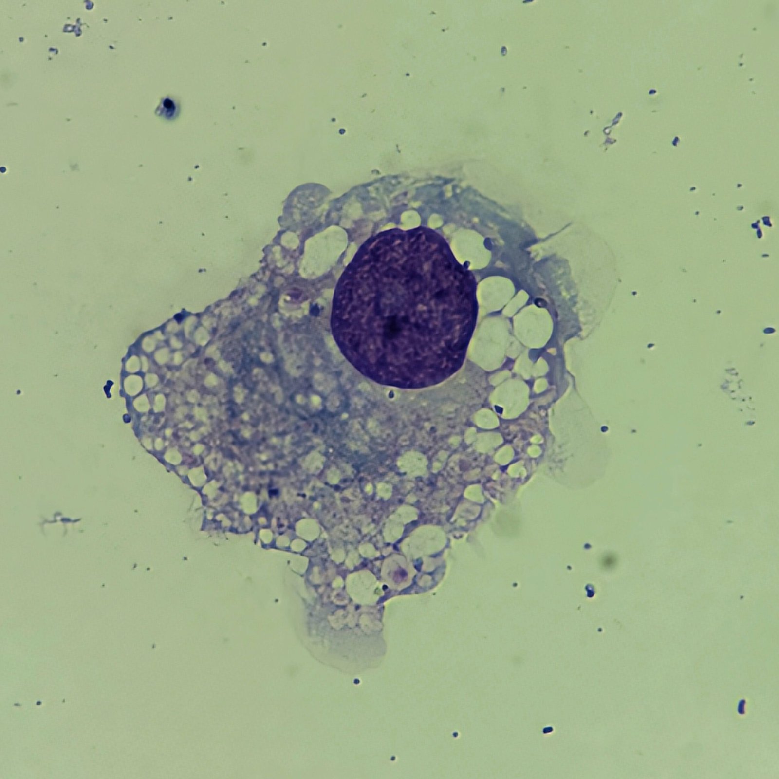
Gallery