Sideroblastic anemia is defined by the presence of ringed sideroblasts in the bone marrow. Iron accumulates in the mitochondria of erythroid precursors and can be seen with a Prussian blue iron stain as blue granules around at least one third of the nucleus. In peripheral blood, red blood cells with pappenheimer bodies can be seen, known as siderocytes. The prefix for these cells comes from the Greek word for iron: “sidero”.
As opposed to iron deficiency anemia, ferritin and iron levels will be increased.
This anemia may be congenital or acquired. Mean Corpuscular Volume (MCV) of the red cells will vary depending on the cause.
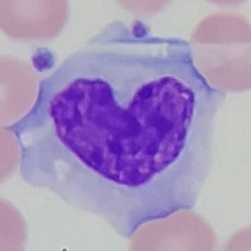
Image of ringed sideroblasts from imagebank.hematology.org.
Congenital
Hereditary sideroblastic anemia is caused by genetic defects affecting mitochondrial heme synthesis. Iron continues to build up in the mitochondria without proper utilization which causes the ringed sideroblasts and iron overload. This usually causes a microcytic, hypochromic anemia.
Acquired
Primary sideroblastic anemia can develop as a form of myelodysplastic syndrome (MDS).
Secondary sideroblastic anemia can develop from a number of causes and is reversible when the underlying issue is treated. Deficiencies in vitamin B6 or copper can cause this anemia as they are important cofactors in heme synthesis. Zinc toxicity is another cause, as it reduces the absorption of copper and can lead to a copper deficiency. Other causes include heavy metal poisoning, certain drugs, or alcoholism.
Acquired sideroblastic anemia usually causes a normocytic or macrocytic anemia.
References
- 4.3: Sideroblastic Anemia – Medicine LibreTexts
- Hematology and Oncology – Merck Manual Professional Edition
- Causes, Symptoms & Treatment
- NCBI Bookshelf
- What Are Siderocytes and What Do They Indicate? – ScienceInsights
- Ring Sideroblasts: Formation and Associated Conditions – Biology Insights
- Zinc Toxicity: Understanding the Limits – PMC