Blister and bite cells are most often caused by removal of Heinz bodies or other erythrocyte inclusions by the spleen. Blister cells can also be caused by fibrin strands rupturing the red cell membrane. Bite cells can form from these blistered red blood cells.
These cells are seen in G-6-PD deficiency and hemoglobinopathies.
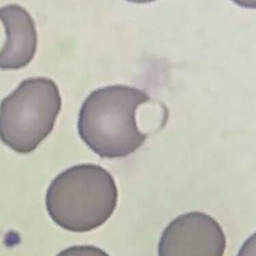
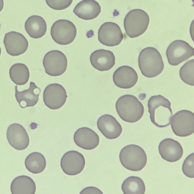
Blister Cell Appearance
Blister cells have a vacuole on one side of the cell.
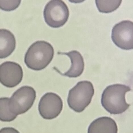
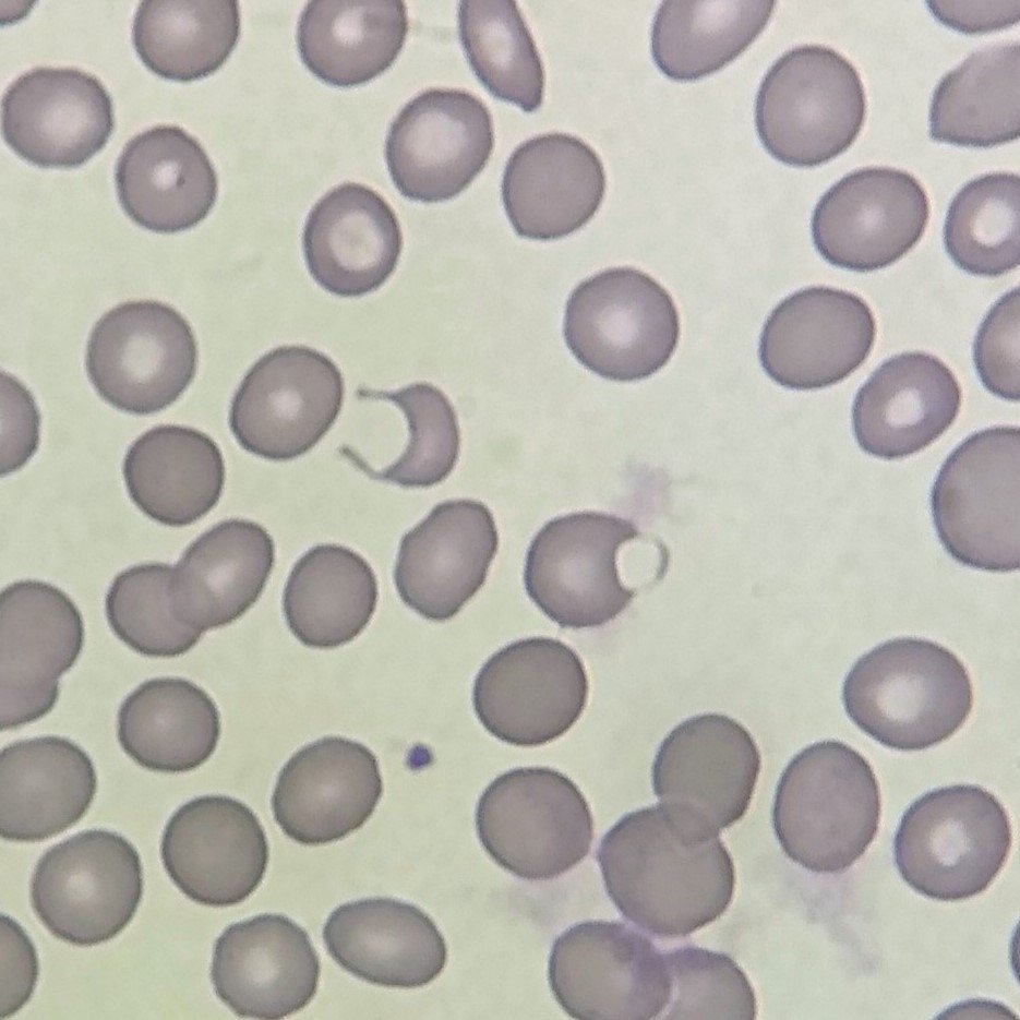
Bite Cell Appearance
Bite cells are indented and look like someone has taken a bite out of it.
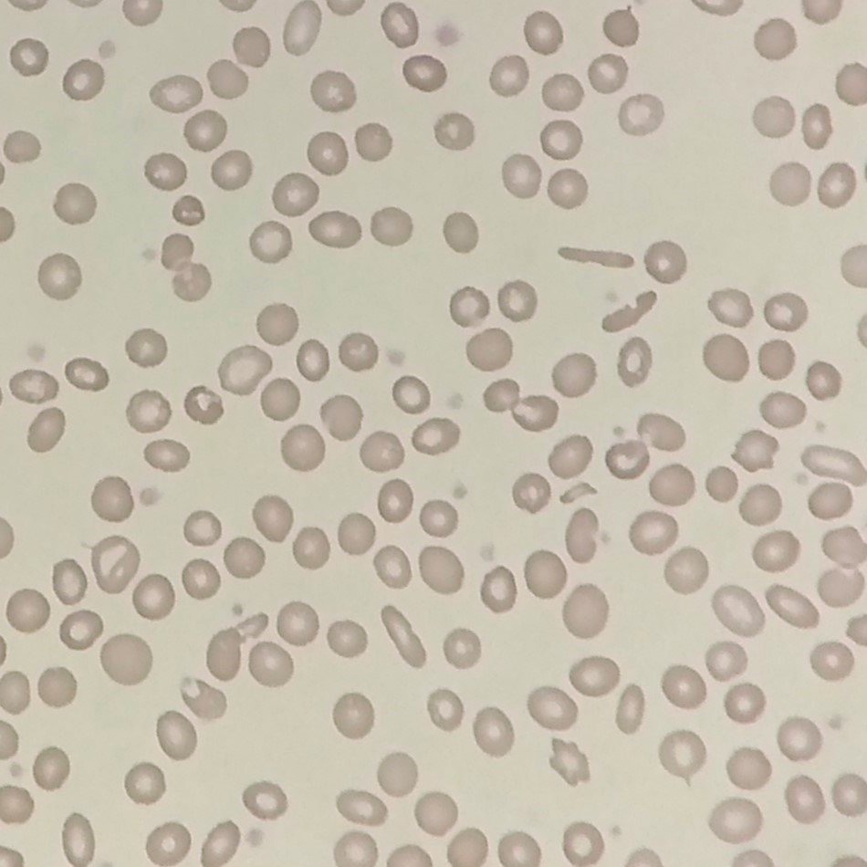
Lookalikes
Residual water on a slide can affect the red blood cell membrane, making it look pitted. However, these pits are much smaller than the indents seen in bite cells. The slide should remade as the artifact can obscure true morphological abnormalities.
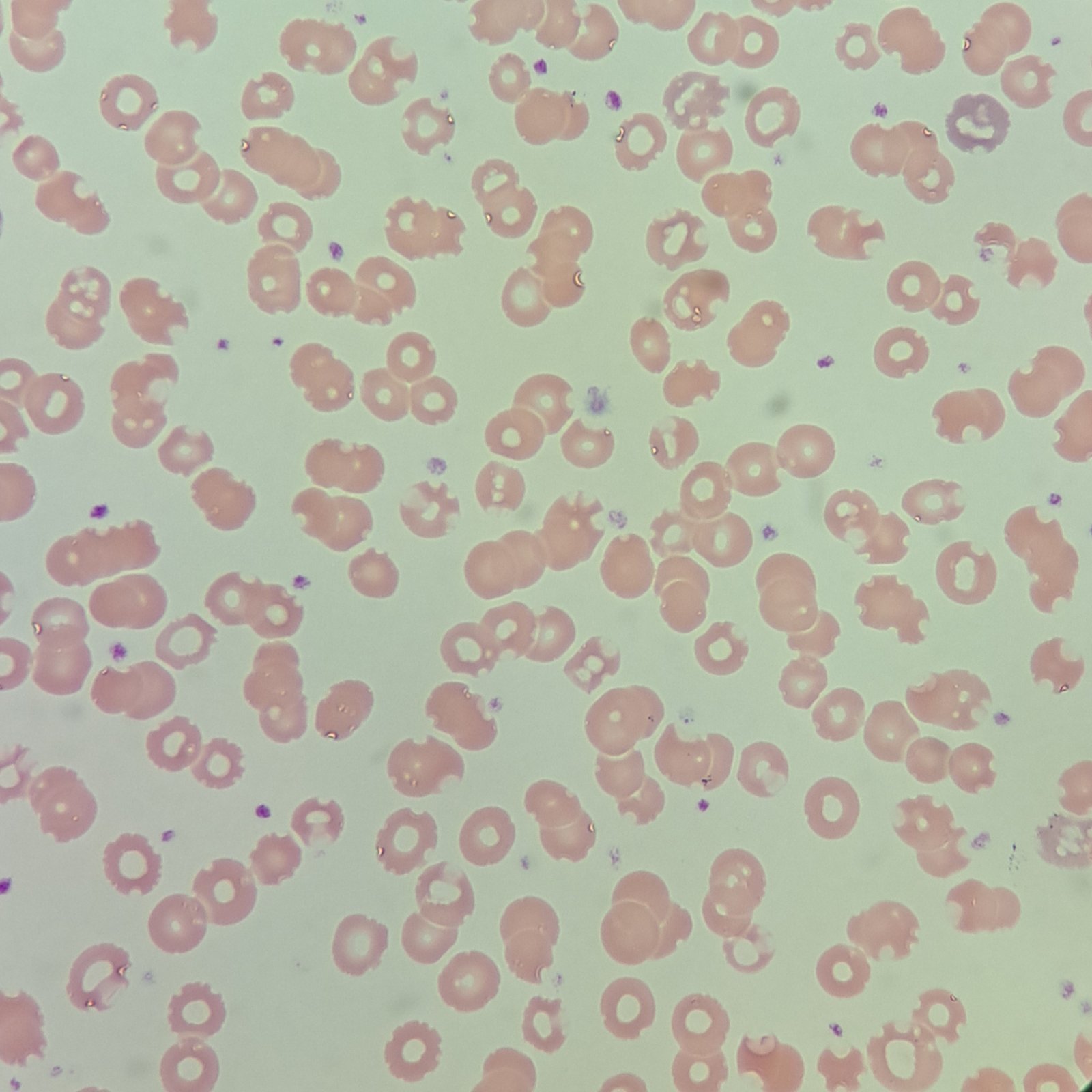
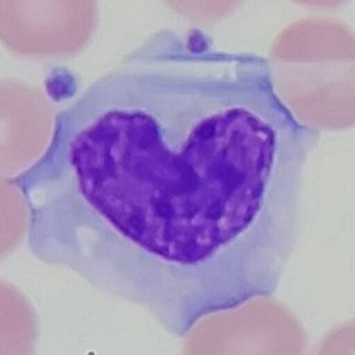
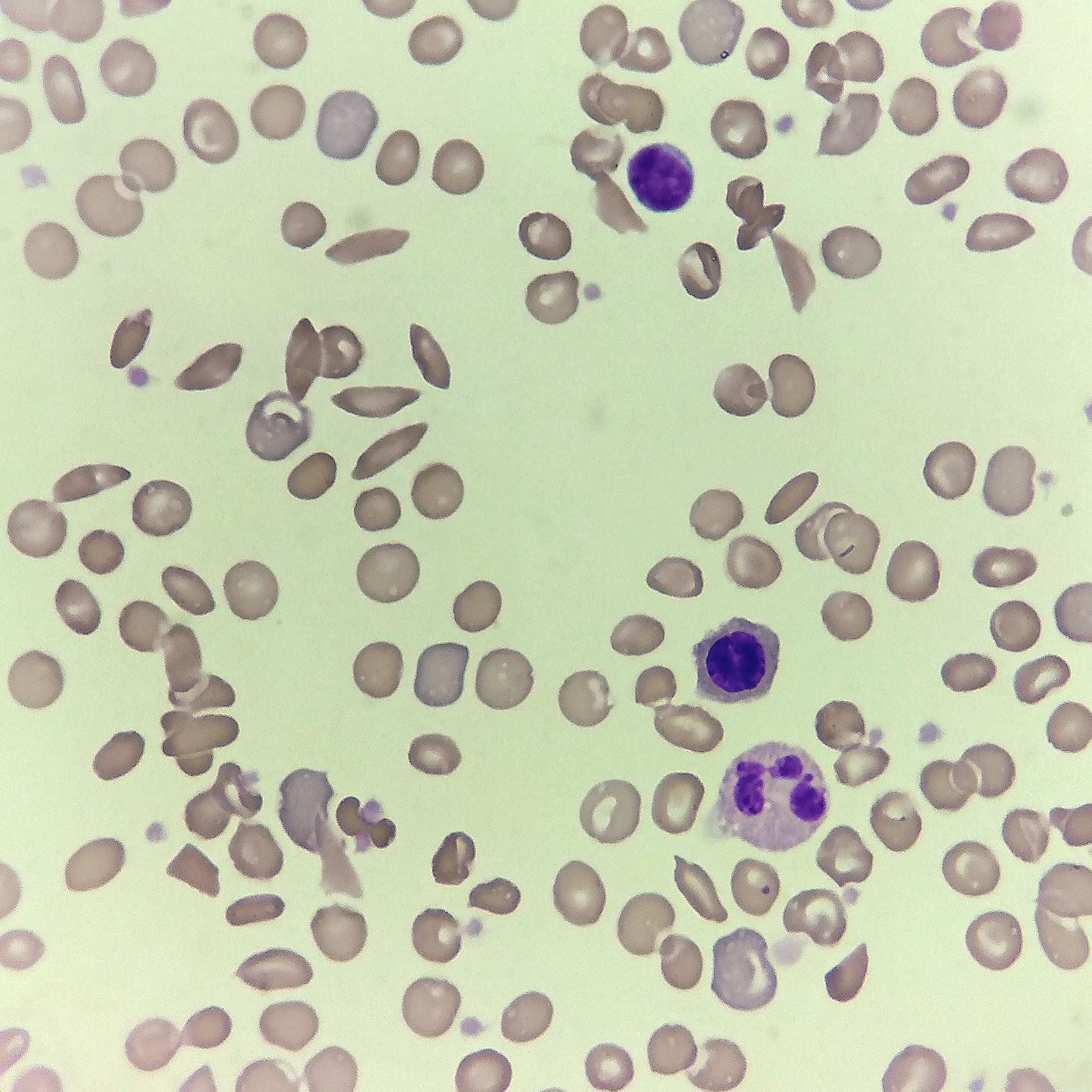
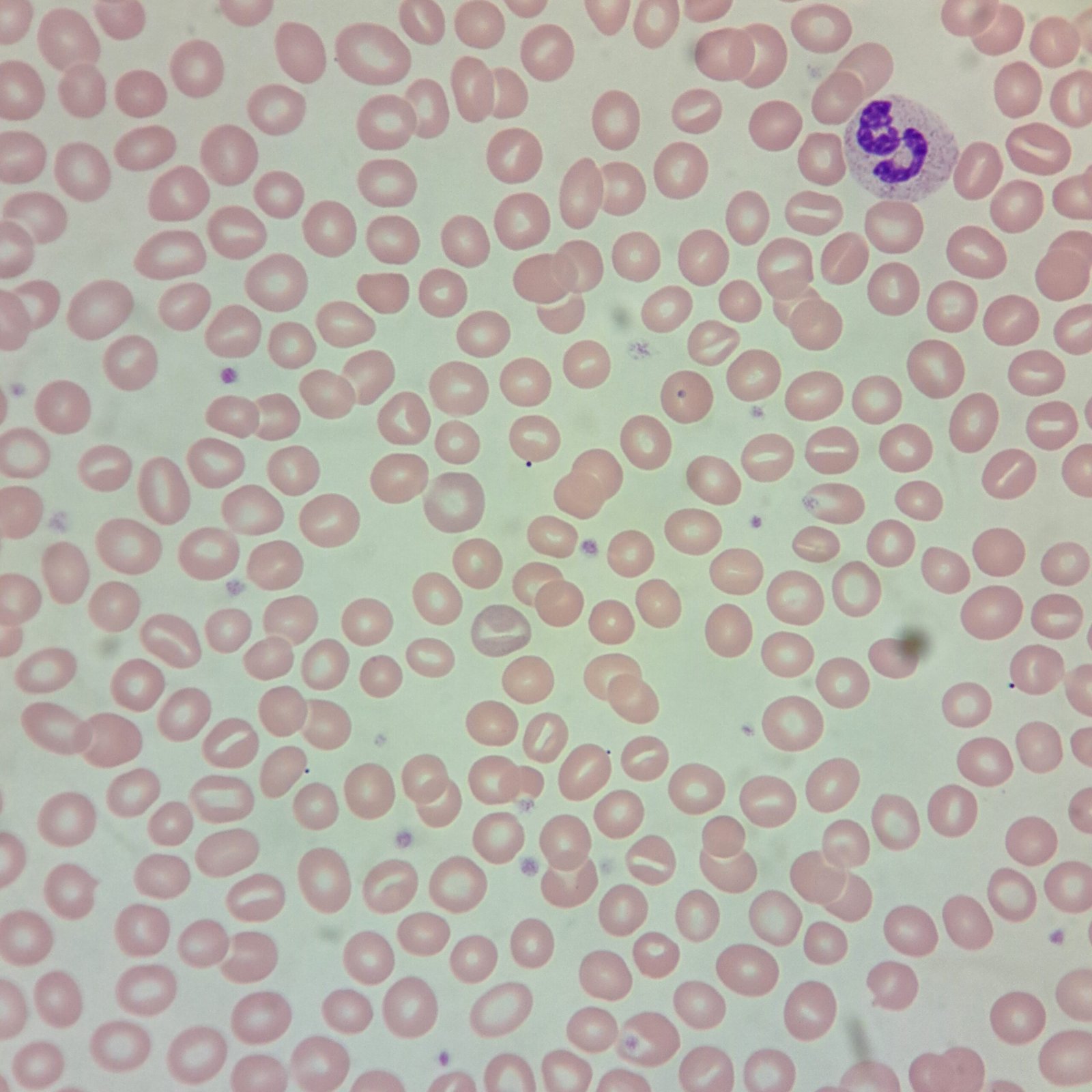
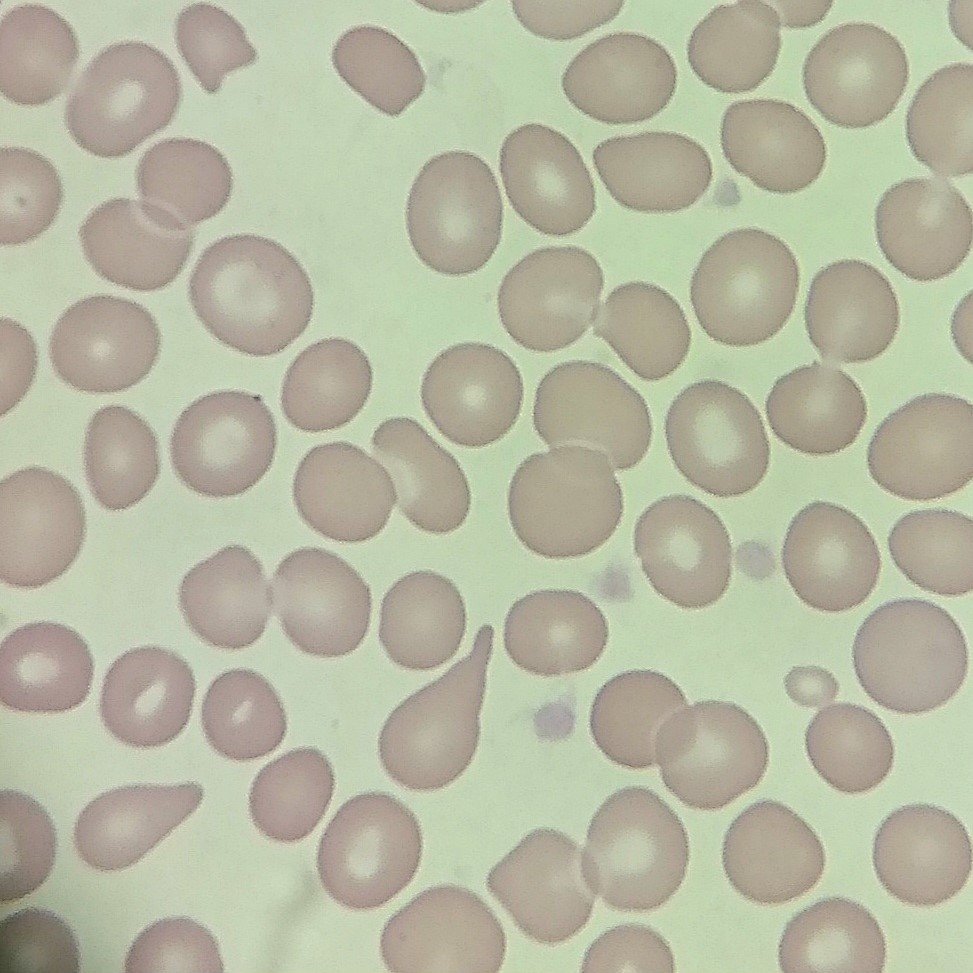
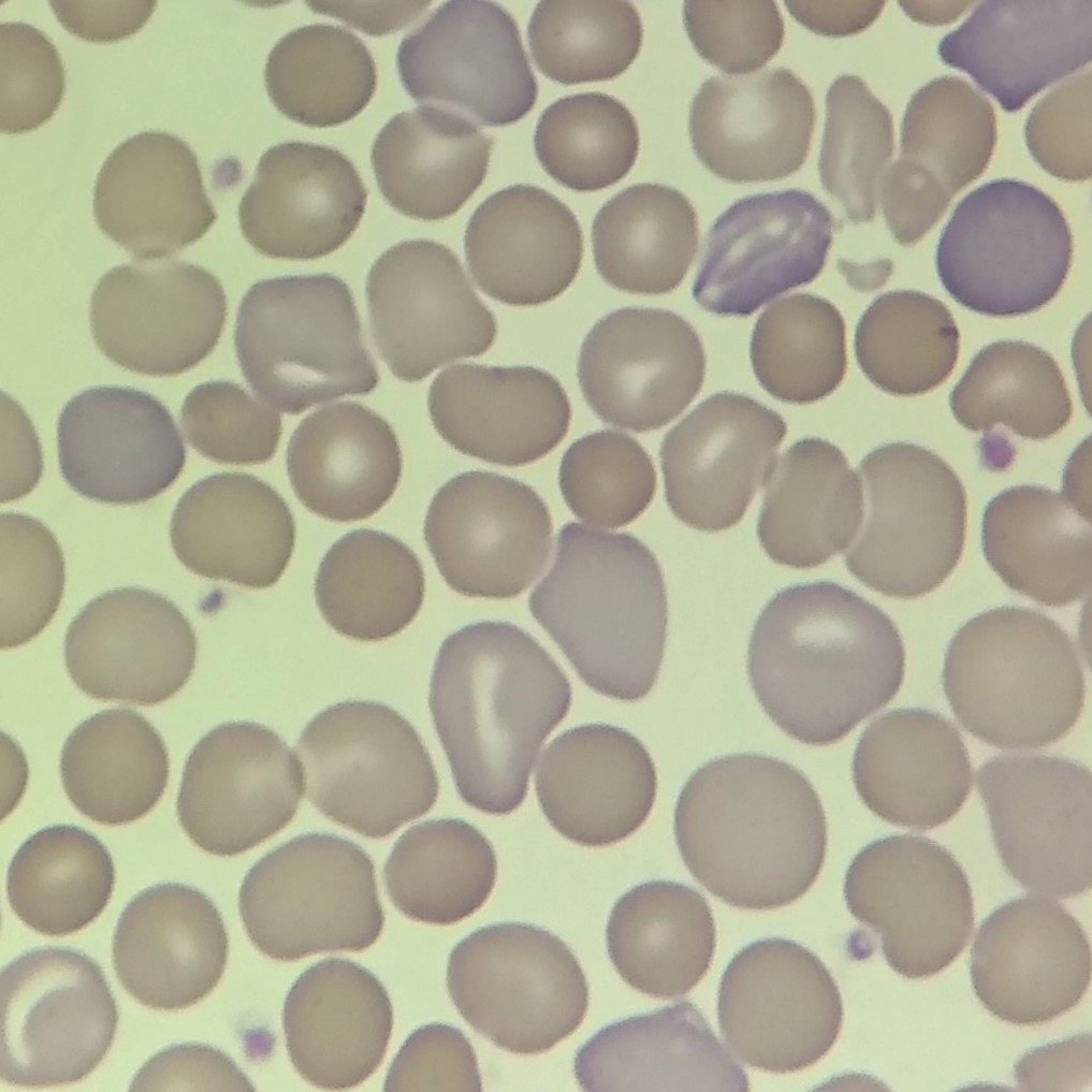
Gallery of Blister & Bite Cells


References
Authored by Rachel Harper, Medical Laboratory Scientist (ASCP)
Last reviewed: January 2026
For educational and reference purposes only, this is not medical advice.